A Radiologist’s Role: More Than Just Reading Scans

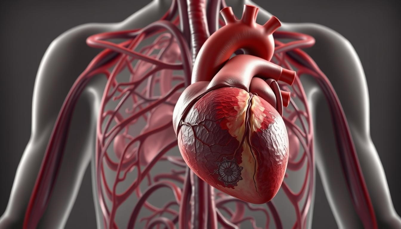
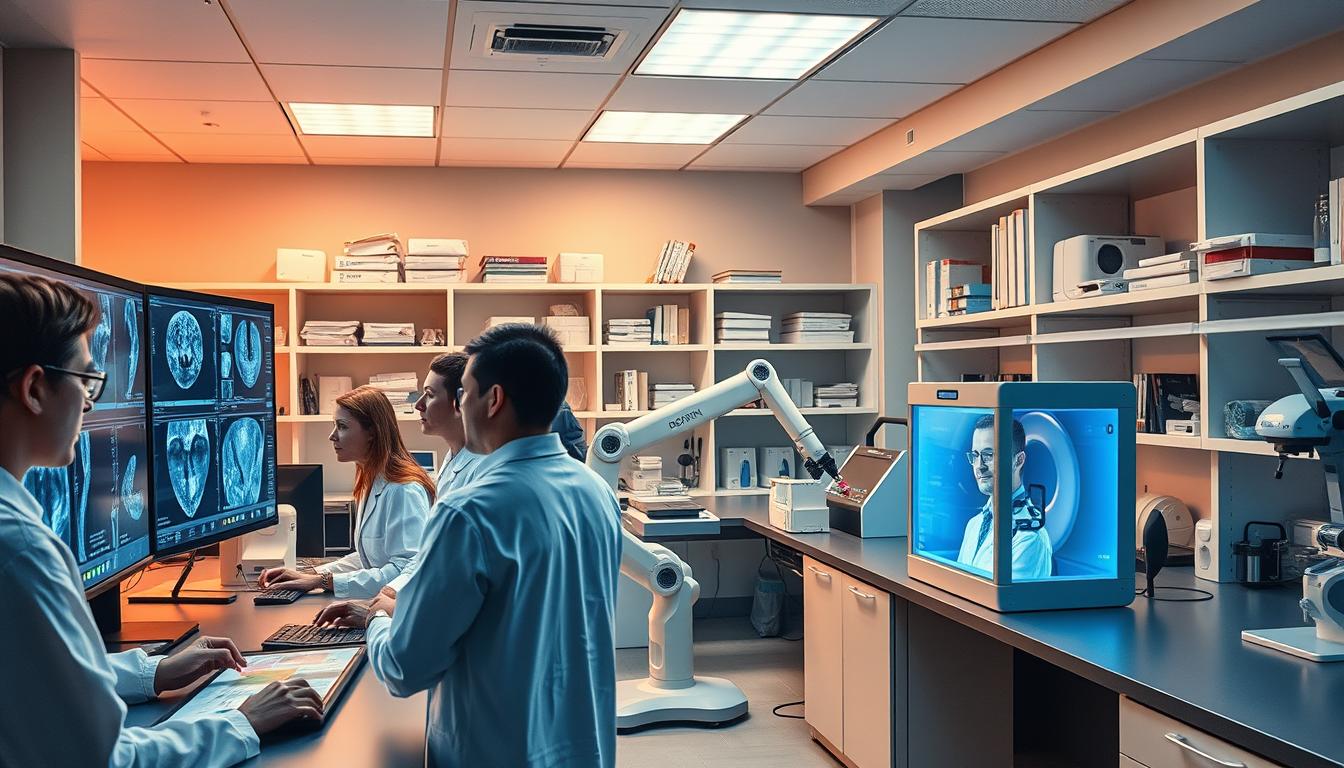
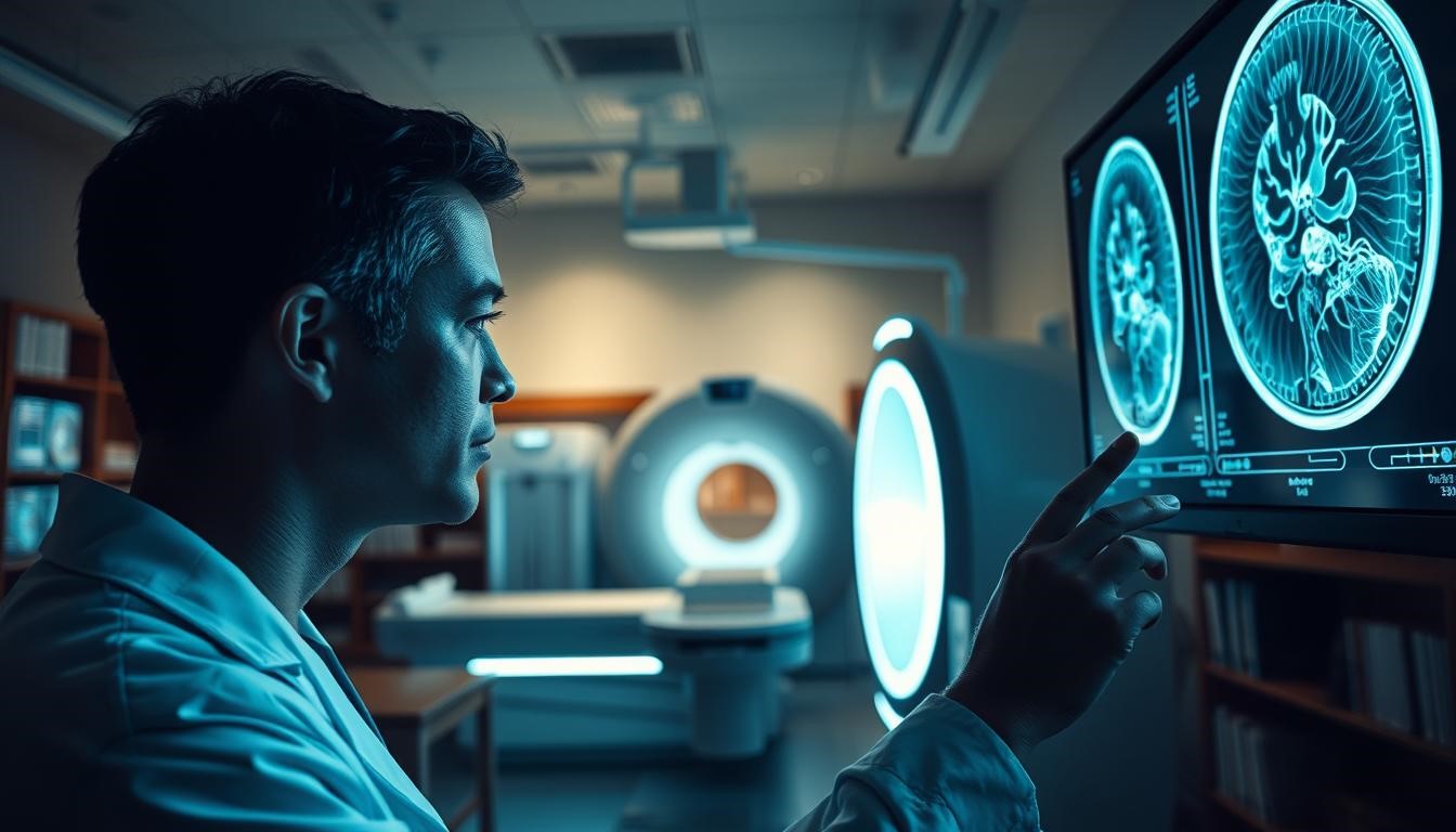
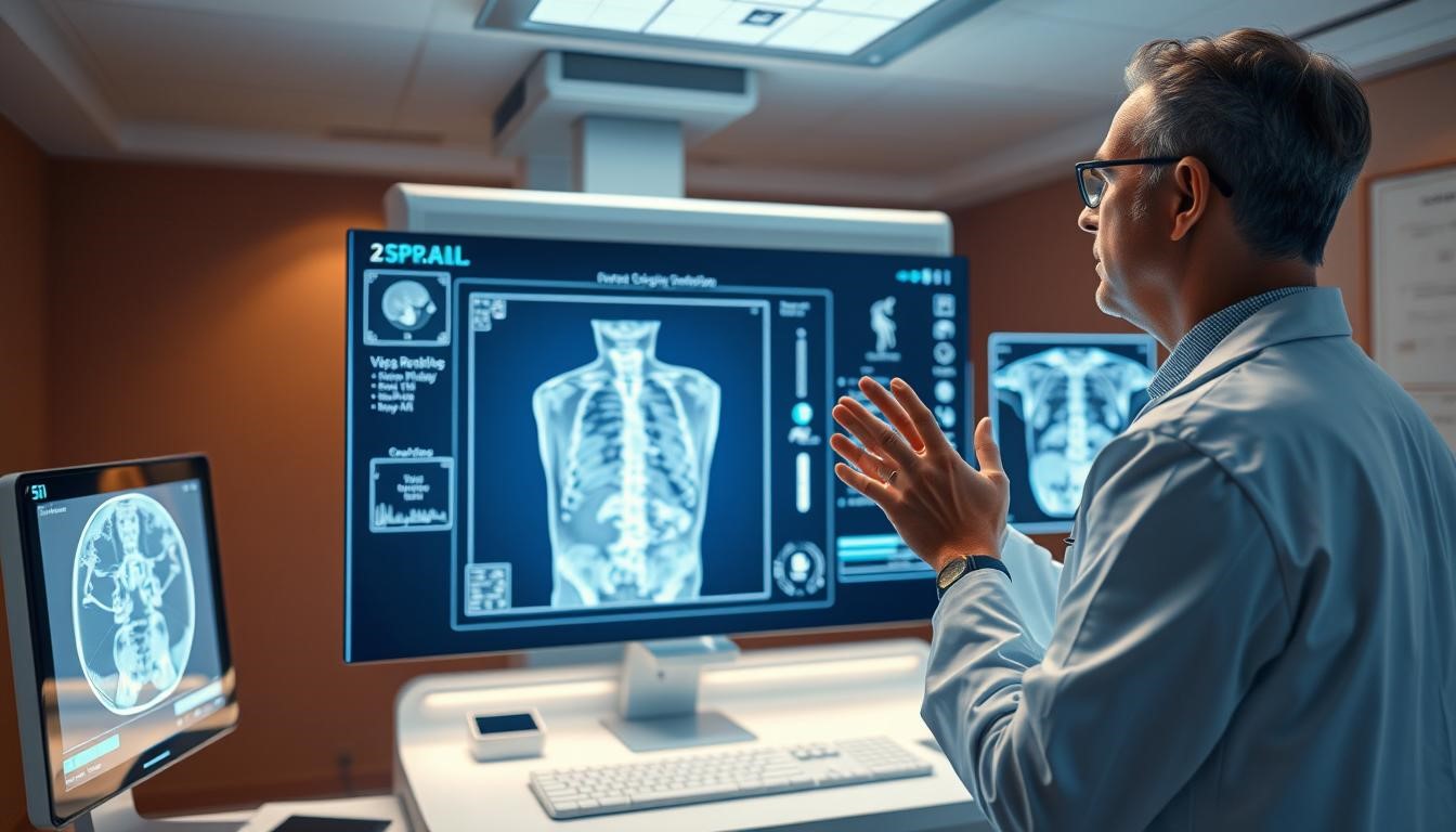
In healthcare, Medical Imaging is key for diagnosing and treating patients. At the center are Radiologists, experts in reading imaging scans.

Radiologists do more than just look at scans. They are key to patient care, working with others to find the right diagnosis and treatment.

Thanks to their knowledge in Radiology, they find the root of symptoms. This helps healthcare teams make the best decisions for patients. It shows how important Radiologists are in today’s healthcare.

medical workflows , Radiology with AI, Artificial Intelligence

What is a Radiologist?

Radiologists are medical experts who use imaging tech to diagnose and treat diseases. They are key in healthcare, interpreting images to guide treatments.

Medical Imaging Specialists

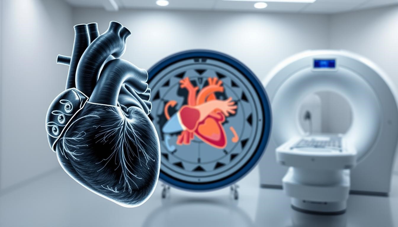
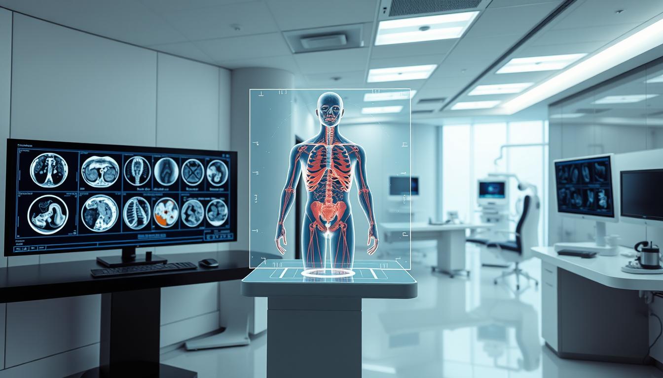
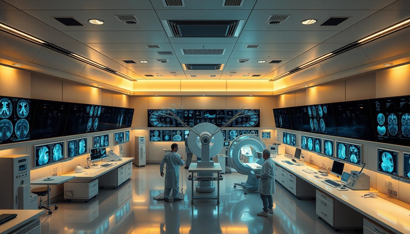
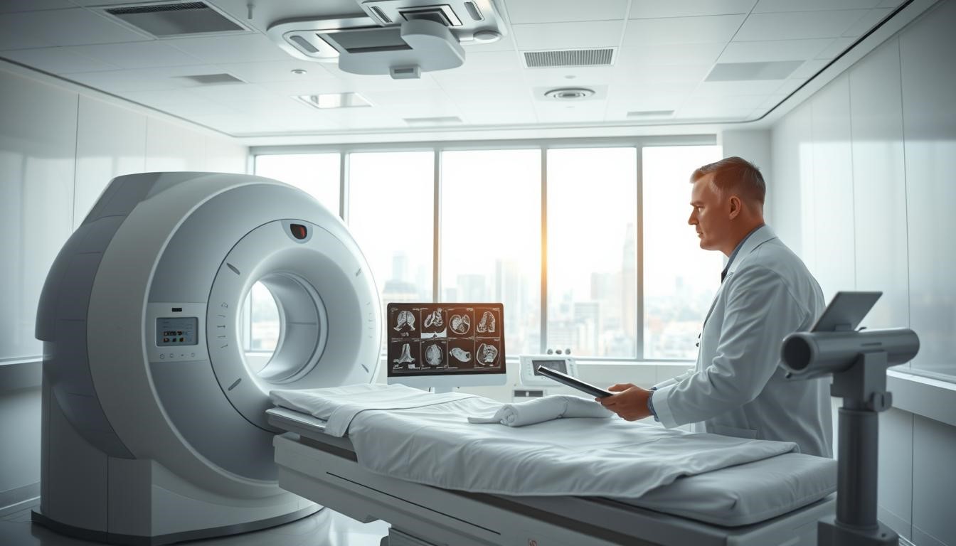
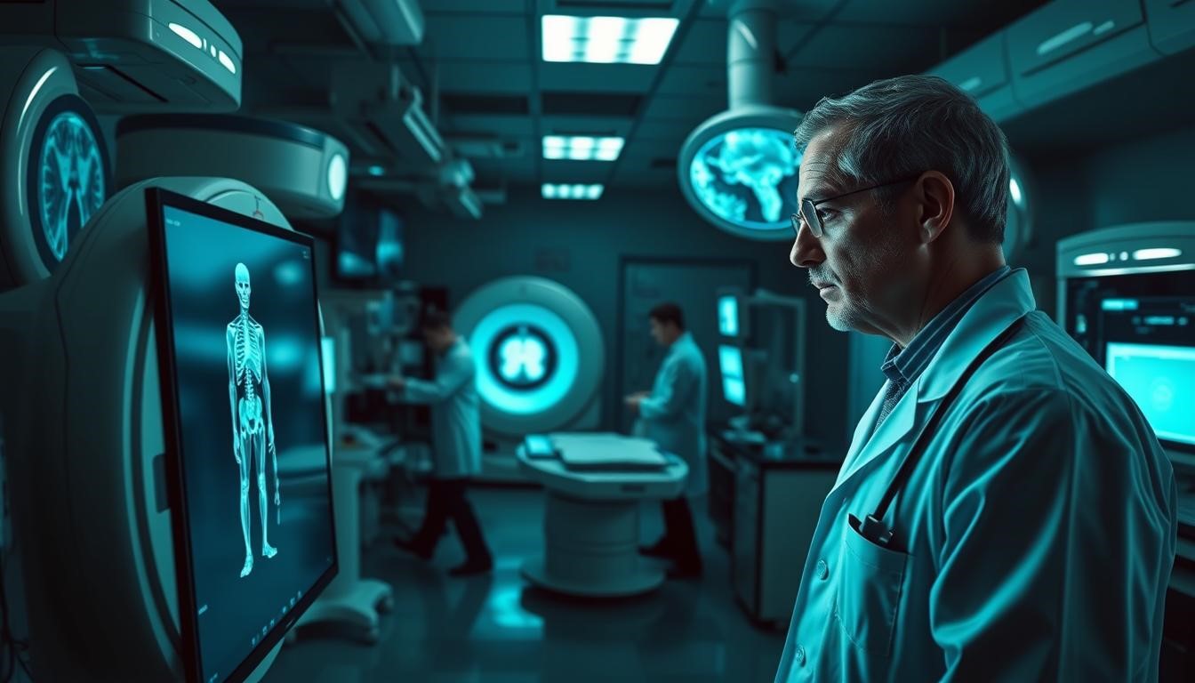
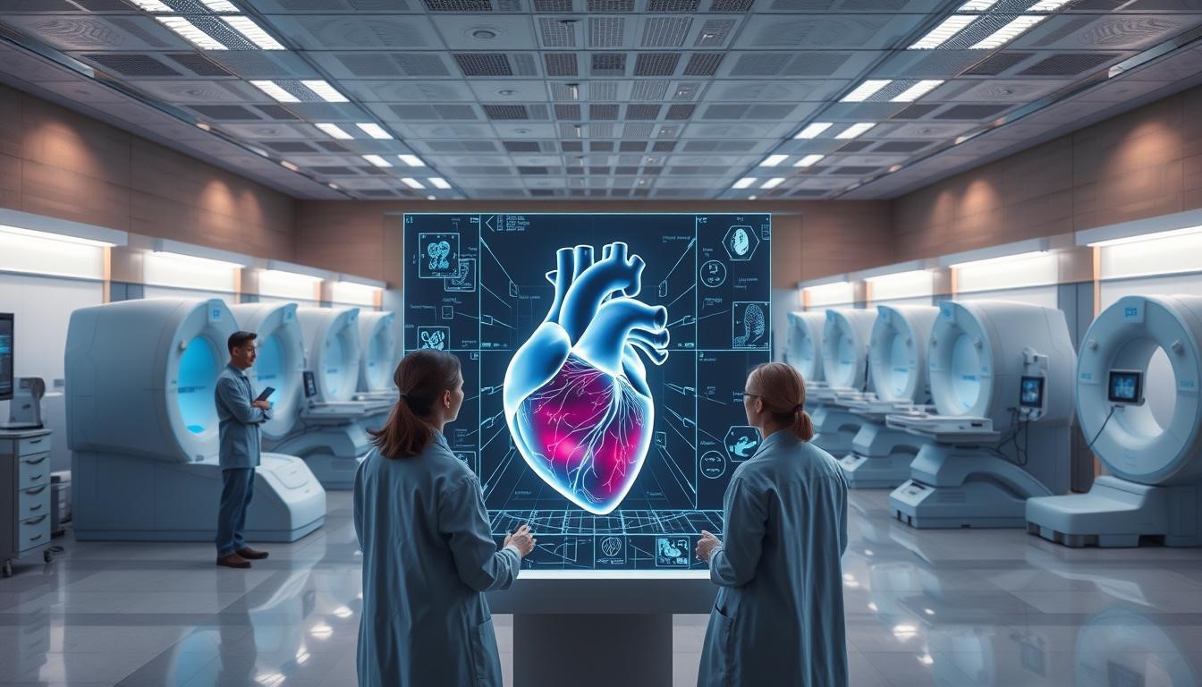
Radiologists are skilled in diagnostic imaging. They use X-rays, ultrasound, CT, MRI, and PET scans to see inside the body.

Education and Training Requirements

To become a radiologist, you need lots of education and training. This takes years after high school.

Medical School and Residency

First, you study for four years in medical school to get an MD or DO. Then, you do a four-year residency in radiology.

Fellowship and Specialization

After residency, many radiologists get more training in fellowships. These last one to two years and focus on special areas like neuroradiology or pediatric radiology.

The Radiologist’s Core Clinical Responsibilities

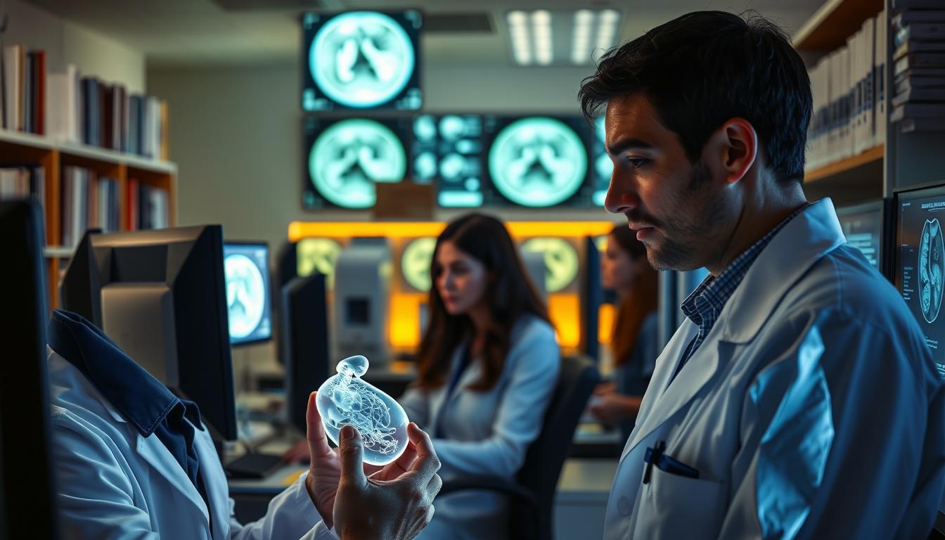
Diagnostic imaging is key to a radiologist’s job. They interpret medical images to diagnose and treat diseases.

Diagnostic Image Interpretation

Radiologists are key in diagnosing conditions. They use X-rays, CT scans, and MRI images. Their skills help spot problems and guide treatment.

Accurate image interpretation is vital for patient care. It affects treatment decisions and outcomes.

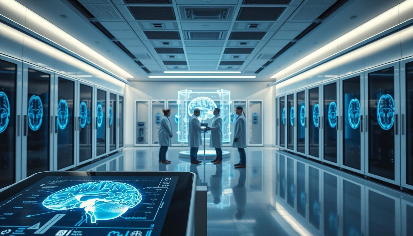
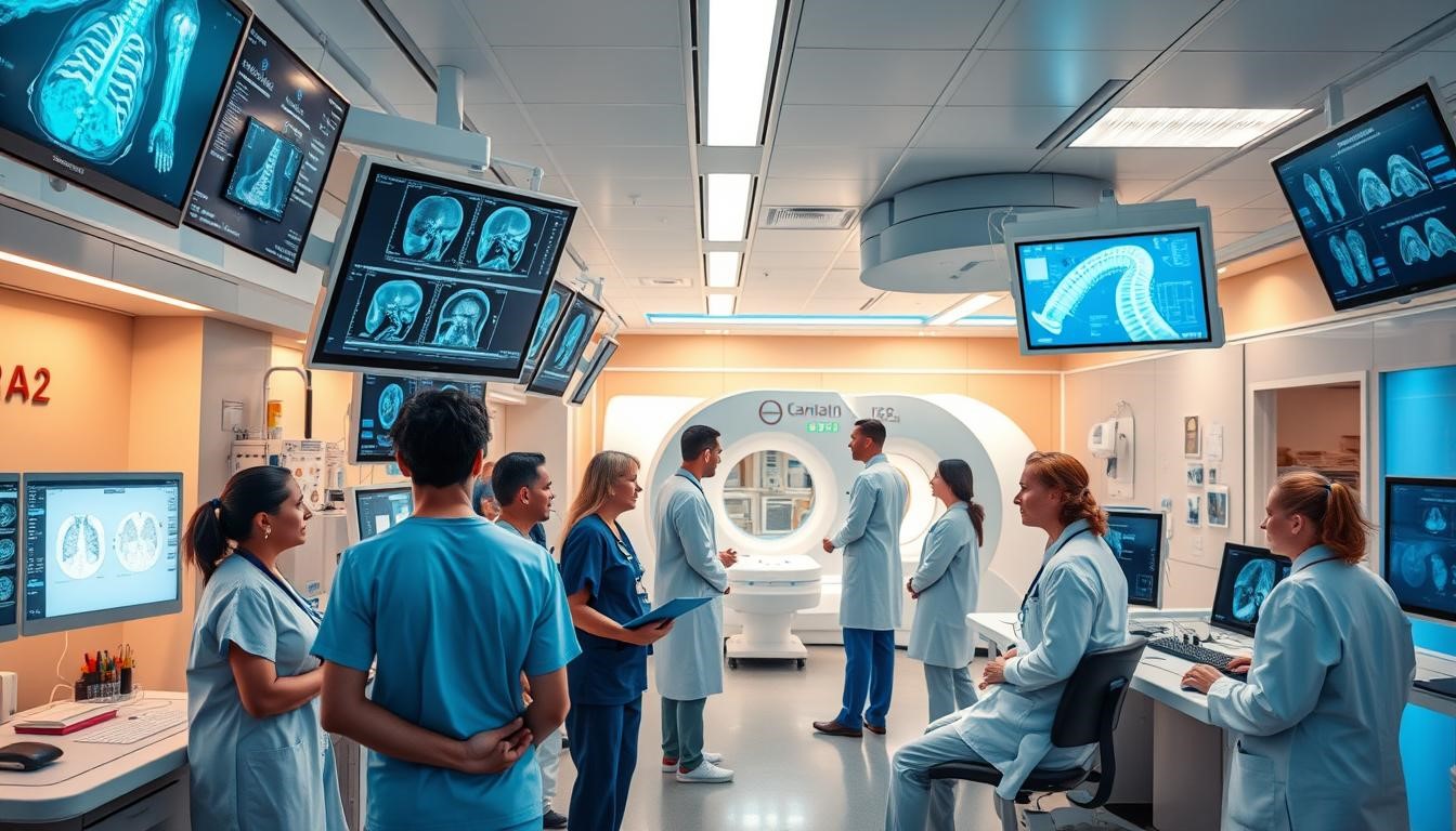
Consultation with Healthcare Teams

Radiologists work with healthcare teams for complete care. Their input is essential in multidisciplinary team discussions.

Multidisciplinary Tumour Boards

In tumour boards, radiologists share their expertise. They help discuss patient cases and plan treatments. Their insights cover all aspects of a patient’s condition.

Emergency Department Collaboration

Radiologists work with emergency staff to quickly diagnose and manage acute conditions. Their fast image interpretation is critical in emergencies.

Radiologists combine image interpretation with teamwork for patient care. Their work is vital for accurate diagnoses and effective treatments.

Beyond Image Interpretation: A Radiologist’s Extended Role

Radiologists do more than just look at images. They also perform procedures and care for patients directly. Their skills greatly improve patient outcomes, making them key in healthcare.

Interventional Procedures and Treatments

Interventional radiology is a big part of a radiologist’s job. It involves doing minimally invasive procedures to diagnose and treat diseases. These can include things like angioplasty, stenting, biopsies, and tumor treatments.

By using imaging, radiologists can do these procedures with great accuracy. This means less need for open surgery and quicker recovery times for patients.

Direct Patient Care and Communication

Good patient care is not just about technical skills. It also needs compassionate communication. Radiologists talk to patients, explain procedures, discuss results, and answer their questions.

Explaining Procedures and Results

It’s important to communicate clearly about medical procedures and results. Radiologists make sure patients understand their conditions and treatments. This helps create a supportive environment where patients feel free to ask questions.

Managing Patient Anxiety

Many patients get anxious before medical procedures. Radiologists help by reassuring them, explaining what will happen, and making sure they’re comfortable. This caring approach makes the experience better for patients.

Radiologist Subspecialties and Areas of Expertise

Radiology is a wide field with many subspecialties. These areas are key in patient care. They let radiologists focus on specific imaging needs, improving diagnosis and treatment.

The subspecialties in radiology are diverse and complex. Some main areas include:

• Neuroradiology: Focuses on imaging of the brain, spine, and nervous system.

• Musculoskeletal Radiology: Deals with imaging of muscles, bones, and joints.

• Pediatric Imaging: Involves imaging for diagnosis and treatment of children.

Neuroradiology and Brain Imaging

Neuroradiologists specialize in diagnosing brain and nervous system conditions. They use MRI and CT scans. Their skills are vital for managing stroke, tumors, and neurological disorders.

Musculoskeletal and Sports Medicine Radiology

Musculoskeletal radiologists diagnose injuries and conditions in muscles, bones, and joints. They use imaging to guide treatments, mainly in sports medicine.

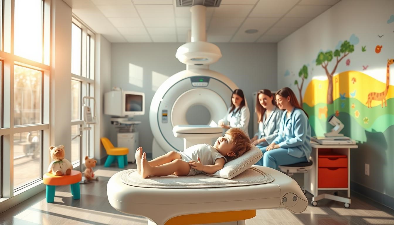
Pediatric and Women’s Imaging

Pediatric radiologists are trained for imaging children, considering their unique anatomy and physiology. Women’s imaging includes mammography and breast imaging for female patients.

These subspecialties show the wide range of radiology. They also highlight the specialized training radiologists get for top-notch patient care.

Challenges Faced by Modern Radiologists

Modern radiologists deal with many challenges every day. The field of radiology is changing fast. New Medical Technology brings both chances and hurdles.

Technological Advancements and Continuous Learning

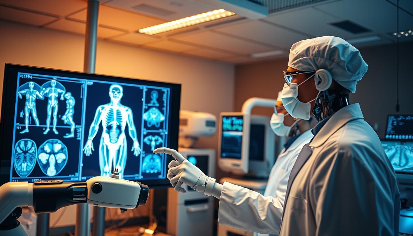
Technological changes in radiology are happening quickly. New tools like AI and machine learning are changing how we diagnose. Radiologists need to keep learning to use these new tools well.

Workload Management and Preventing Burnout

Managing work is key for radiologists to stay healthy and give great care. There are two main things to focus on:

• Handling more imaging studies, which means finding better ways to work.

• Creating a balance between work and life to avoid burnout.

Increasing Imaging Volume

More people need imaging tests as they get older. Radiologists must find ways to handle this increase. They can use technology to make their work more efficient.

Work-Life Balance Strategies

It’s important for radiologists to have a good balance between work and personal life. This can be done by having flexible hours, wellness programs, and clear boundaries between work and personal time.

By tackling these challenges, radiologists can make their work better. They can also improve patient care and keep themselves healthy in a tough healthcare world.

The Future of Radiology: Emerging Trends and Technologies

The future of radiology is changing fast. New trends and technologies are making a big difference. These changes will affect how radiologists work and the care they give.

Diagnostic Innovations

Artificial Intelligence (AI) is becoming a big help in radiology. AI can look at images faster and more accurately. This means radiologists can make better diagnoses quicker.

Teleradiology and Global Practice

Teleradiology is also changing radiology. It lets radiologists read images from anywhere. This opens up new ways to practice radiology worldwide.

Remote Consultations

Remote consultations are becoming more common. Radiologists can now give advice from afar. This makes patient care better and cuts down on the need for face-to-face meetings.

Addressing Healthcare Disparities

Teleradiology is also tackling healthcare gaps. It brings radiology services to places that need them most. This helps improve health outcomes and reduce care disparities.


Radiologists do more than just look at medical images. They are key in patient care, working with others to find the right diagnosis and treatment. Their skills are vital for helping patients get better.

Radiology keeps growing with new tech like artificial intelligence and teleradiology. Radiologists need to keep up with these changes. This helps them improve patient care and make things run smoother in hospitals.

Radiologists are essential to healthcare, giving insights that guide treatment plans. They use their knowledge and care for patients to make healthcare better. Their work is a big part of improving health outcomes.

FAQ

What is the role of a radiologist in healthcare?

Radiologists are key in healthcare, doing more than just reading scans. They work with other doctors to give accurate diagnoses and treatment plans.

What kind of education and training do radiologists receive?

Radiologists get a lot of education and training. They finish medical school, then do a residency and sometimes a fellowship.

How do radiologists contribute to patient care?

Radiologists help by reading images, doing procedures, and talking to patients and doctors.

What are some of the subspecialties within radiology?

Radiology has many subspecialties like neuroradiology and women’s imaging. Each needs special training and skills.

How do radiologists stay current with technological advancements?

Radiologists keep up with new tech by always learning. This includes using artificial intelligence in their work.

What is teleradiology, and how is it changing radiology practice?

Teleradiology lets radiologists read images from anywhere. It’s making radiology services more accessible and changing how radiologists work.

How do radiologists manage patient anxiety during procedures?

Radiologists and their teams help patients feel less anxious. They explain things clearly, offer support, and make sure patients are comfortable.

What is the significance of multidisciplinary tumour boards?

Multidisciplinary tumour boards are important. They bring together doctors, including radiologists, to plan treatments for complex cases. This shows how vital radiologists are in cancer care.


The Silent Killer: How Imaging Helps Detect and Manage Heart Disease

Cardiac health is a big worry all over the world. Heart disease is a top reason for deaths.

Imaging tech is key in spotting and handling this issue. It shows us how the heart works and looks.

Healthcare experts use advanced imaging to find problems early. This means they can act fast to help patients.

This approach not only makes care better. It also shows how vital it is to keep the heart healthy. This is done with the help of technology and smart healthcare.

Heart Disease: The Silent Threat to Your Health

Heart disease is a silent threat because it often goes unnoticed until symptoms are severe. It’s important to know the types of heart conditions, why they’re often missed, and the risk factors. This knowledge helps keep your heart healthy.

Common Types of Cardiovascular Conditions

Cardiovascular disease includes many heart and blood vessel conditions. These include coronary artery disease, heart failure, and arrhythmias. Each can be serious if not managed well.

Why It Often Goes Undetected Until Too Late

Heart disease often starts quietly, with symptoms showing up only when it’s too late. This is because the heart tries to keep working even when it’s damaged. Regular check-ups and knowing the signs are key to catching it early.

Risk Factors Everyone Should Know

Several factors increase your risk of heart disease. These include high blood pressure, high cholesterol, smoking, diabetes, and a family history of heart disease. Knowing these risks helps you take steps to lower your own risk.

Understanding heart disease and its risks helps protect your heart. Regular health checks and a healthy lifestyle are key to preventing heart disease.

The Evolution of Cardiac Imaging Technology

Cardiac imaging technology has changed a lot, making it easier to diagnose heart disease. We’ve moved from old methods to new digital ones. This change has greatly improved how we find and treat heart problems.

From X-rays to Advanced Digital Imaging

At first, X-rays were used to look at the heart. But they didn’t show much. Then, new tech like angiography came along. It let doctors see the heart’s blood vessels better.

Now, we have digital tools like CT scans, MRI, and echocardiography. These help doctors get clear pictures of the heart. This makes diagnosing heart disease more accurate.

How Modern Technology Has Improved Diagnosis

Switching to digital imaging has made diagnosing heart disease much better. With MRI and CT scans, doctors can see the heart’s details clearly. This helps find heart problems early and plan treatments better.

Thanks to modern cardiac imaging, patients are getting better care. This has really improved their health outcomes.

Key Imaging Techniques That Save Lives

Heart disease diagnosis and treatment have greatly improved thanks to new imaging techniques. These tools help doctors see the heart’s details clearly. This leads to better diagnoses and treatment plans.

Echocardiography: Ultrasound for the Heart

Echocardiography, or an echo, uses sound waves to show the heart’s images. It’s a safe and painless test. It helps doctors see the heart’s parts and how they work. Echocardiography is great for finding heart valve issues and heart failure.

Cardiac CT Scans: Detailed Heart Anatomy

Cardiac CT scans make detailed X-ray images of the heart. They’re good for spotting blockages in the heart’s arteries. They help doctors see the heart’s details, like finding coronary artery disease.

Cardiac MRI: Complete Heart Check

Cardiac MRI uses a strong magnetic field and radio waves to show the heart’s images. It checks the heart’s structure and function. It’s great for looking at the heart’s work and finding issues like cardiomyopathy.

Nuclear Cardiac Imaging: Function and Blood Flow

Nuclear cardiac imaging uses tiny amounts of radioactive material to see the heart’s function and blood flow. It’s good for spotting coronary artery disease and checking the heart’s overall health. It gives doctors important info on the heart’s blood flow and function, helping them plan treatments.

These imaging methods have changed how we diagnose and treat heart disease. They help doctors give more accurate diagnoses and effective treatments. By knowing each method’s strengths, doctors can pick the best one for each patient.

How Imaging Transforms Heart Disease Management

Advanced imaging technologies have changed how we manage heart disease. They give detailed views of the heart’s structure and function. This helps doctors diagnose and treat heart disease better.

Early Detection and Prevention Strategies

Imaging is key in catching heart disease early. It lets doctors act fast and prevent serious problems. Tools like echocardiography and cardiac CT scans spot issues before symptoms show up.

Treatment Planning and Monitoring Progress

After finding heart disease, imaging helps plan treatments and check how they’re working. Cardiac MRI and nuclear cardiac imaging show how well the heart is working. This lets doctors adjust treatments as needed.

Guiding Minimally Invasive Procedures

Imaging also helps with less invasive procedures, like transcatheter aortic valve replacement (TAVR). It gives doctors real-time images. This makes complex procedures more precise and safe.

Imaging has greatly improved heart disease management. It has led to better patient outcomes and saved many lives. As these technologies get better, we’ll see even more tailored care for heart disease patients.

Making Cardiac Imaging Work for You

Exploring cardiac imaging might seem tough, but it’s key to your heart health. It’s more than just a test; it opens the door to understanding your heart. This knowledge helps you make better choices for your care.

When to Seek Imaging: Warning Signs You Shouldn’t Ignore

Knowing when to get cardiac imaging is important. Signs like chest pain, shortness of breath, or palpitations are serious. If you notice these, talk to your doctor about getting an imaging test.

Navigating Insurance Coverage and Costs

Knowing your insurance is vital for cardiac imaging. Many plans cover these tests, but coverage varies. Always check your policy and talk about costs with your doctor.

Questions to Ask Your Healthcare Provider

When talking to your doctor about imaging, be ready with questions. Ask about the type of imaging they suggest, the risks and benefits, and how it will affect your treatment.

Being proactive and informed helps you use cardiac imaging to your advantage. This is a big step towards better heart health.

Taking Control of Your Heart Health

Knowing how imaging helps find and manage heart disease is key to keeping your heart healthy. Advanced cardiac imaging lets people take steps to prevent and manage heart conditions.

Being aware of risk factors and warning signs is important. Regular check-ups and screenings can spot problems early. This means doctors can act quickly.

Staying informed and working with your healthcare team is vital. You can learn how to keep your heart in top shape. This includes understanding insurance, treatment options, and asking questions for the best care.

FAQs

What is heart disease and how does it affect the body?

Heart disease is when the heart’s structure and function are affected. This can lead to serious issues like heart failure, arrhythmias, and heart attacks.

What are the common types of cardiovascular conditions associated with heart disease?

Common types include coronary artery disease, heart failure, arrhythmias, and valvular heart disease. These can greatly affect a person’s life and increase their risk of death.

Why is heart disease often referred to as a “silent killer”?

Heart disease often doesn’t show symptoms until a major event, like a heart attack. It’s important for people to know their risk factors and get regular check-ups.

What are the key risk factors for developing heart disease?

Key risk factors include high blood pressure, high cholesterol, diabetes, smoking, obesity, family history, and a sedentary lifestyle. These can be managed through lifestyle changes and medical treatments.

How has cardiac imaging technology evolved over time?

Cardiac imaging has moved from traditional X-rays to advanced digital methods. This includes echocardiography, cardiac CT scans, cardiac MRI, and nuclear cardiac imaging. These advancements have greatly improved diagnosis.

What is echocardiography and how is it used in heart disease diagnosis?

Echocardiography uses ultrasound to provide detailed information about the heart. It helps diagnose and manage various heart conditions.

How do cardiac CT scans contribute to heart disease diagnosis?

Cardiac CT scans give detailed images of the heart’s anatomy. They help healthcare providers assess coronary artery disease and other heart conditions.

What are the benefits of cardiac MRI in assessing heart health?

Cardiac MRI offers a detailed look at the heart’s structure and function. It helps diagnose and manage a wide range of heart conditions effectively.

When should I seek cardiac imaging, and what are the warning signs I shouldn’t ignore?

Seek cardiac imaging if you have symptoms like chest pain, shortness of breath, or dizziness. Or if you have risk factors for heart disease. Early detection is key for effective management.

How can I navigate insurance coverage and costs associated with cardiac imaging?

Talk to your healthcare and insurance providers to understand coverage and costs. This will help you find the most suitable and affordable imaging options.

What questions should I ask my healthcare provider about cardiac imaging?

Ask about the benefits and risks of different imaging techniques. Also, ask about the procedure and how the results will guide your treatment. This ensures you get the best care.


Pediatric Radiology: Gentle Imaging for Our Youngest Patients

PediPediatric imaging needs a special touch to help children. Gentle Imaging Techniques are key in Child Imaging. They help get accurate diagnoses without causing too much pain or risk.

Imaging kids is different from adults. It requires special methods and tools. These are designed to meet the unique needs of young patients, ensuring they get the best care.

This article will dive into why pediatric radiology is so important. We’ll look at the challenges of imaging children and the advanced methods used to tackle them. It will give a full picture of this vital field.

The Unique Challenges of Pediatric Imaging

Imaging children is not just about making adult procedures smaller. It needs a special approach. Pediatric radiology requires knowing the unique features of children’s bodies and minds.

Anatomical and Developmental Differences

Children’s bodies grow and change a lot. Their anatomy is different from adults. Smaller size, changing bone density, and growing organs need special imaging methods. It’s also important to use lower radiation doses to protect them.

Psychological Considerations When Imaging Children

Imaging can scare children, causing them to feel anxious. Radiologists must think about how imaging affects children’s minds. They should use simple language, reassure them, and involve parents. This helps make the experience less scary.

Building Trust with Young Patients and Their Families

Building trust is key for successful pediatric imaging. Radiologists and staff need to understand and support children and their families. They should explain things clearly and offer emotional support. This helps reduce anxiety and makes the process smoother.

Specialized Radiology Techniques for Pediatric Patients

Specialized radiology techniques are key in pediatric care. They offer gentle and accurate imaging solutions. These methods tackle the unique challenges of imaging children, including anatomical and developmental differences, and psychological considerations.

Child-Specific Imaging Protocols

Pediatric radiology departments create child-specific imaging protocols. These ensure diagnostic imaging is safe and effective for young patients. The protocols take into account the child’s age, size, and the clinical question being addressed.

Tailored imaging protocols reduce the need for repeat exams. This lowers radiation exposure and boosts diagnostic accuracy.

Pediatric radiology imaging

Radiation Dose Management and Safety

Managing radiation dose is vital in pediatric radiology. Techniques like adjusting exposure settings on radiography and CT equipment are key. Using alternative imaging modalities like ultrasound or MRI when needed also helps minimize radiation exposure.

• Implementing ALARA (As Low As Reasonably Achievable) principles guides radiation dose management.

• Regularly maintaining and calibrating equipment ensures optimal performance.

• Training staff on radiation safety and dose optimization is essential.

Equipment Modifications for Pediatric Applications

Pediatric imaging needs equipment designed for children’s smaller size. Modifications include using size-appropriate devices and making comfort-enhancing adaptations. These help reduce anxiety and improve image quality.

Size-Appropriate Devices

Using devices designed for pediatric patients ensures safe and effective imaging. This includes pediatric-specific coils for MRI and appropriately sized detectors for radiography.

Comfort-Enhancing Adaptations

Adaptations like pediatric-friendly immobilization devices and comfort measures during imaging are vital. They reduce distress and improve the experience for children and their families.

By using these specialized techniques and equipment modifications, pediatric radiology advances. It provides high-quality diagnostic imaging that meets the unique needs of children.

Common Pediatric Radiology Procedures and Applications

Radiological imaging is key in pediatric care, with several important procedures. These help doctors assess and treat children’s health issues. From fractures and infections to complex birth defects, pediatric radiology is essential.

X-rays for Growth Assessment and Fracture Detection

X-rays are vital in pediatric radiology. They help check bone growth and find fractures. Modern X-rays use low radiation, making them safe for kids.

Ultrasound Benefits in Pediatric Diagnosis

Ultrasound is a top choice in pediatric care. It’s non-invasive and doesn’t use harmful radiation. It’s great for looking at organs, checking the heart, and guiding treatments. Ultrasound’s live images are perfect for kids, who can move around.

CT and MRI: Indications and Considerations

CT and MRI scans give detailed views of inside the body. They’re key for complex diagnoses. CT scans are quick and detailed, while MRI shows soft tissues well without radiation. Choosing between them depends on the child’s size and the specific question.

Nuclear Medicine Studies for Children

Nuclear medicine uses tiny amounts of radioactive tracers. In kids, it helps check organ function, find infections, and spot cancers. The tracers are chosen to keep radiation low while getting important info.

Interventional Radiology in Pediatric Care

Interventional radiology uses imaging to guide small procedures. In kids, it can include biopsies, drainages, and vascular work. It’s a less invasive option than surgery, helping kids recover faster.

Creating Child-Friendly Imaging Environments

A well-designed imaging environment greatly improves care for kids. It makes the experience better and helps get accurate results.

Physical Space Design and Atmosphere

The space for imaging should be designed for kids. Use bright colors, fun decor, and games to keep them distracted.

Key elements of a child-friendly imaging environment include:

• Comfortable seating for families

• Toys and entertainment for children

• Clear explanations and preparation materials

Age-Appropriate Preparation Techniques

It’s important to prepare kids for imaging in a way they can understand. Explain it simply, use pictures, or even practice with mock scans.

Parental Involvement During Procedures

Having parents there can comfort a child. But, parents need to know their role and how to support their child.

Sedation and Anesthesia Approaches

Sometimes, sedation or anesthesia is needed for the child’s comfort and the procedure’s success.

When Sedation Is Necessary

Sedation is used when a child can’t stay calm or is too nervous. The choice depends on the child’s health, age, and the imaging needed.

Minimizing Risks and Recovery

When sedation or anesthesia is used, it’s key to watch the child closely. Have emergency plans ready. Afterward, watch for any bad effects and help with recovery.


Pediatric radiology has made great strides in providing safe and effective imaging for kids. Looking ahead, we see more advancements in Pediatric Radiology Advances. These will help improve how we diagnose and treat young patients.

By using imaging methods made for kids, managing radiation, and making places friendly for children, radiologists can reduce stress. New technologies and techniques will also help. This means better care for kids, tailored to their needs.

As we move forward, focusing on the well-being of children is key. This approach ensures Pediatric Radiology Advances meet kids’ needs. It leads to better health and a better life for them.

FAQ

What is pediatric radiology?

Pediatric radiology focuses on imaging and diagnosing children. It uses X-rays, ultrasound, CT, and MRI. It covers newborns to teens.

Why is pediatric radiology important?

It’s key because kids’ bodies are growing and different from adults. They need special imaging methods and tools for safe and effective scans.

What are some common pediatric radiology procedures?

Procedures include X-rays for growth and fractures, ultrasound for belly and pelvic issues. CT and MRI scans are for detailed images. Nuclear medicine studies check organ function.

How do radiologists prepare children for imaging procedures?

Radiologists use simple explanations and child-friendly language. They offer emotional support to make kids feel at ease during scans.

What role do parents play during pediatric imaging procedures?

Parents are very important. They help calm their child’s fears and provide reassurance. They also help keep their child steady during the scan.

Are sedation and anesthesia used in pediatric radiology?

Yes, sedation and anesthesia are used when needed. They help kids stay calm and steady during scans. But, they’re used carefully, considering the child’s age and health.

How is radiation exposure managed in pediatric radiology?

Radiation exposure is managed by using special imaging protocols for kids. The goal is to use the least amount of radiation. Non-radiation methods like ultrasound and MRI are also used.

What are some of the challenges faced in pediatric radiology?

Challenges include needing special equipment and techniques. Managing radiation exposure is also a big challenge. Plus, there are unique anatomical and developmental differences in kids, as well as their emotional needs.


Cost-Effective Research Using AI in Radiology

The field of radiology is changing fast, needing cost-effective research methods. Old ways of doing research in radiology take a lot of time and money. They involve looking at data by hand and understanding it.


can change this, making it easier and more accurate to look at big data sets.

AI helps find patterns and trends that humans might miss. This leads to new ideas and discoveries. It can also make care better for patients.

Cost-effective research in radiology

Cost-effective research in radiology is possible with AI. It makes the research process faster and cheaper. This is because it cuts down on the need for manual data analysis.

The Current Challenges in Radiology Research

The field of radiology research faces many challenges. These issues affect different parts of research, like how studies are done and how data is handled.

Traditional Research Methods and Their Limitations

Old ways of doing research in radiology rely on manual analysis. This can be slow and full of mistakes. It also needs a lot of people and resources.


Cost-effective research in radiology

Rising Costs in Medical Imaging Studies

Medical imaging studies are getting more expensive. This makes it hard to do research without going over budget. Higher costs come from needing better technology and dealing with big data and long analysis times.

Data Management Hurdles in Radiological Research

Managing data well is key in radiology research. But, it’s a big challenge. The huge amounts of data need special systems to keep it safe and easy to use. Bad data management can cause problems and even lose data, hurting research results.

To move forward, we need to tackle these challenges. By knowing what’s holding us back, we can find new ways. For example, using AI could make research better, faster, and cheaper.

The Transformative Impact of AI in Radiology

AI is changing radiology a lot. It’s making how we look at medical images better and faster. This change is making diagnoses more accurate and quicker.

Machine Learning Algorithms for Image Analysis

Machine learning is key in AI for radiology, mainly for looking at images. These algorithms can learn from data, spot patterns, and make smart choices without being told what to do.

Supervised vs. Unsupervised Learning Approaches

Supervised learning uses labeled data to train algorithms. They learn from examples and apply this to new data. On the other hand, unsupervised learning finds patterns in data without labels. It uncovers hidden structures or groups.

Neural Networks in Diagnostic Imaging

Neural networks are a big help in medical imaging. They can spot complex patterns in images, like signs of disease.


A sleek, modern medical imaging workstation set against a backdrop of clinical white walls and futuristic technology. In the foreground, an AI-powered algorithm visualizes a 3D holographic model of a human anatomy, with intricate details and precise annotations. The middle ground features various diagnostic imaging modalities, including CT scans, MRI images, and ultrasound displays, all seamlessly integrated with the AI system. The background showcases a panoramic view of a state-of-the-art radiology department, where medical professionals utilize the cutting-edge AI-driven tools to enhance their diagnostic capabilities and optimize patient outcomes. Crisp lighting illuminates the scene, creating a sense of clinical precision and technological innovation.

Current Applications in Clinical Settings

AI is being used more in hospitals to make diagnoses better and faster. It helps in finding problems and making the work flow smoother.

Detection and Classification of Abnormalities

AI helps find and sort out problems in medical images, like tumors or breaks. This can cut down the time to get a diagnosis and help patients more.

Workflow Optimization and Prioritization

AI also makes the work in radiology better by sorting cases by how urgent they are. This means important cases get looked at first.

Validation Methods for AI Algorithms

It’s very important to check if AI algorithms work well and are safe. This means testing them with known data and making sure they work in real situations.

By doing this, doctors can rely on AI to make diagnoses better and faster. This helps take care of patients even more.

Cost-Effectiveness of AI-Powered Radiology Research

AI makes radiology research more cost-effective and improves results. It’s not just a tech upgrade; it’s a smart financial move. It’s changing how we do medical research.

Reducing Time and Resource Requirements

AI cuts down the time and resources needed for radiology research. It does this in a few ways:

  • Automated Image Analysis and Annotation: AI quickly analyzes and annotates images. This used to take a lot of time and effort.
  • Streamlining Research Workflows: AI organizes and manages data. This makes the research process smoother and reduces the workload for researchers.

Improving Diagnostic Accuracy and Efficiency

AI speeds up and improves the quality of research. It brings several benefits:

  1. Reducing False Positives and Negatives: AI is trained to spot abnormalities accurately. This lowers the rate of false positives and negatives.
  2. Enhancing Radiologist Productivity: AI automates routine tasks. This lets radiologists focus on more complex cases, boosting their productivity.

Long-term ROI for Healthcare Institutions

Healthcare institutions see big returns on investment with AI in radiology. They save on labor costs, get better patient outcomes, and open up new research avenues.

Cost Comparison: Traditional vs. AI-Enhanced Research

AI-enhanced research is more cost-effective than traditional methods. It cuts down on manual data analysis, speeds up research, and boosts accuracy. All these factors lower costs.

In summary, AI in radiology research offers many benefits. It saves time and resources, improves accuracy, and boosts productivity. As healthcare adopts more AI, the long-term benefits will be significant.

Implementing AI Solutions for Cost-Effective Research

To use AI for cheaper research, radiology teams need to focus on a few key areas. Adding AI can make research faster and more accurate.

Required Infrastructure and Technology

A strong setup is key for AI in radiology. You’ll need high-performance computing hardware and advanced data storage solutions. Also, having high-quality imaging equipment is important. Make sure it works well with what you already have.

Training and Integration Strategies

Good training is vital for AI success. Radiology staff should learn about AI and how it works. A smart plan for adding AI to your work is also important.

  • Develop a tailored training program for radiology staff
  • Establish clear guidelines for AI algorithm validation
  • Monitor and adjust AI system performance regularly

Case Studies: Successful Implementation Models

Many hospitals have made AI work in radiology research. For example, a top hospital cut image analysis time a lot. Another place used AI to boost patient results.

Ethical Considerations and Regulatory Compliance

AI in radiology raises big ethical questions. Keeping patient data private and following rules is critical. Hospitals must follow FDA guidelines and be open about their AI use.

Conclusion

AI has changed radiology, making research and diagnosis cheaper. It uses machine learning and data analysis to work faster and more accurately. This means better results and less time spent on tasks.

Using AI in radiology brings many benefits. It cuts costs, improves patient care, and boosts research speed. As more healthcare places use AI, the savings and improvements will grow. This will lead to more innovation in the field.

AI makes radiology research more efficient and affordable. This leads to better care for patients. The use of AI in radiology is exciting and full of promise for the future.

FAQ

What are the primary benefits of using AI in radiology research?

AI in radiology research boosts accuracy and saves time. It also makes radiologists more productive. This leads to cheaper research.

How does AI improve diagnostic accuracy in radiology?

AI uses machine learning to analyze images. It cuts down on errors and finds problems faster.

What infrastructure and technology are required to implement AI solutions in radiology research?

For AI in radiology, you need strong computers and advanced software. Also, good data storage and integration with health records and image systems.

How can AI be validated for use in radiological research?

AI is tested and checked against human experts. It’s evaluated with metrics like accuracy. It must follow rules too.

What are the possible cost savings of using AI in radiology research?

AI can save money by automating tasks and needing less resources. This makes research more efficient and cost-effective for hospitals.

How does AI impact the role of radiologists in research?

AI helps radiologists by doing routine tasks. It gives them tools for decision-making. This lets them focus on more complex cases.

What are the key considerations for implementing AI solutions in radiology research?

Important things include ensuring data quality and following rules. Also, training staff and keeping an eye on AI performance.

Can AI be used with existing radiology research workflows?

Yes, AI can fit into current workflows. It makes things more efficient. It also opens up chances to improve and change workflows.

Addressing the Radiologist Shortage with AI-Powered Solutions

The shortage of radiologists is a big problem. It affects how well healthcare works and the care patients get. With more need for imaging tests, the lack of radiologists is getting worse.

AI-powered solutions are being looked at to solve this issue. By adding AI to radiology, we can make things run smoother. This also makes tests more accurate and helps radiologists do less work.

This change could really improve how radiology works. It could make patient care better and make things run more smoothly. With so few radiologists, using AI in radiology is a big help.

medical imaging , Machine learning in healthcare

The Growing Radiologist Shortage Crisis in Canada

The shortage of radiologists is a big problem that needs quick action. This issue is not the same everywhere in the country. It changes a lot from one area to another.

Current Statistics and Trends in Canadian Healthcare

Recent numbers show a big shortage of radiologists. This affects how well healthcare works. The country’s healthcare system is struggling to keep up with the needed staff.

It looks like more people need radiologists than there are. This means current staff have to work harder and feel more stressed.

Regional Disparities Across Provinces

There are big differences in how many radiologists each province has. Some places have much fewer than others. For example, rural areas often can’t get to radiology services as easily as cities can.

Impact on Patient Wait Times and Diagnostic Accuracy

The lack of radiologists makes patients wait longer for tests and treatments. This delay can affect how quickly they get help. Also, the extra work can make it harder for radiologists to be accurate.

This shows we really need new ways to solve the shortage of radiologists. We must find ways to improve care for patients.

Understanding AI in Radiology: Core Technologies

AI is changing radiology with machine learning, deep learning, computer vision, and natural language processing. These technologies are making it easier to analyze and understand medical images.

medical imaging , Machine learning in healthcare

Machine Learning and Deep Learning Applications

Machine learning and deep learning are key for analyzing images in radiology. They help create models that learn from big datasets, spot patterns, and predict outcomes. Deep learning, a part of machine learning, uses neural networks to handle complex data like medical images.

Computer Vision in Medical Imaging Analysis

Computer vision is essential for medical image analysis. It lets computers understand and interpret visual data from images. This tech is used to find abnormalities, segment images, and classify diseases.

Natural Language Processing for Radiology Reports

Natural Language Processing (NLP) is used to read and understand radiology reports. It extracts important information and offers insights for diagnosis and treatment planning. NLP helps automate report generation and boosts the accuracy of radiology reports.

How AI-Powered Solutions Address Workforce Challenges

AI is changing radiology by solving big workforce problems. It automates simple tasks and boosts how well doctors can diagnose. This helps reduce the stress on radiologists.

Workflow Optimization and Efficiency Gains

AI makes radiology departments work better. Automated image analysis and preprocessing are key to this improvement.

Automated Image Analysis and Preprocessing

AI quickly goes through lots of images, finding important details. It flags these for doctors to check right away. This makes diagnosis faster and more accurate.

Triage and Prioritization Capabilities

AI can sort and prioritize radiology cases. It looks at how urgent and complex they are. This means critical cases get attention quickly, helping patients and cutting wait times.

  • AI can analyze imaging data to identify critical findings.
  • Prioritization based on AI analysis helps in managing workload.
  • Enhanced patient care through timely intervention.

Reducing Radiologist Burnout and Improving Job Satisfaction

AI takes over routine tasks, cutting down on radiologist burnout. This lets radiologists work on harder cases, making their job more rewarding.

AI is making a big difference in radiology. It’s making workflows better, improving how accurate diagnoses are, and making radiologists happier at work.

Key AI Radiology Applications in Canadian Clinical Practice

AI is changing radiology in world in many ways. It makes diagnoses more accurate, makes work flow better, and helps patients get better care.

Diagnostic Support Systems for Common Conditions

Diagnostic support systems are becoming more common. They use machine learning algorithms to look at medical images. This gives radiologists important information to help them diagnose.

Chest X-ray and CT Scan Analysis

AI is key in analyzing chest X-rays and CT scans. It can spot things like pneumonia or lung nodules early. This means doctors can act fast.

Automated Screening Tools for Early Detection

Automated screening tools are also important. They help find diseases like breast cancer early. This is through analyzing mammograms.

  • Improved detection rates
  • Reduced false positives
  • Enhanced patient care

Quality Assurance and Error Reduction Mechanisms

AI is also vital for quality and reducing mistakes in radiology. It looks at imaging data to find errors or problems. This makes diagnoses more accurate.

Quality assurance mechanisms include checks for image quality and following protocols. Error reduction involves AI finding issues that might be missed by humans.

Implementation Challenges and Considerations

AI is changing radiology, but there are hurdles to overcome for it to work well in Canadian healthcare.

Health Canada Regulatory and Approval Pathways

Getting AI radiology solutions approved by Health Canada is key. You need to know the regulatory approval process. This means showing that your AI is safe and works well.

Following Health Canada’s rules is vital to get your product on the market.

  • Understanding the classification of AI software as a medical device
  • Preparing for pre-market submission requirements
  • Ensuring post-market surveillance and vigilance

medical imaging , Machine learning in healthcare

Integration with Existing Provincial Healthcare Systems

AI solutions need to fit into current healthcare systems well. This means healthcare integration that makes things work smoothly. It’s important for provinces and AI developers to work together.

  1. Developing standardized data exchange protocols
  2. Training healthcare professionals on AI system usage
  3. Monitoring and evaluating AI system performance

Data Privacy and Security Under Canadian Regulations

Data privacy and security are top priorities for AI in radiology. Canadian laws, like PIPEDA, protect patient data. AI providers must keep this data safe.

Overcoming these challenges is essential for AI to improve patient care in Canada. It must meet all the rules.

Canadian Success Stories: AI in Radiology Transforming Healthcare

AI is changing radiology in Canada. Hospitals and startups are leading the way. They are making healthcare better with AI.

Hospital Implementation Case Studies

In Toronto, a hospital used AI to help doctors. It made diagnosis faster and more accurate. They saw a 30% drop in wait times and a 25% cut in mistakes.

Measurable Outcomes and Benefits

AI in radiology has brought big wins. It has made patient care and hospital work better. A study showed AI helped radiology teams work more efficiently.

Canadian AI Radiology Startups and Research Innovations

Canada has a growing number of AI radiology startups. They are pushing the limits of radiology. These startups are creating new tools and checks, helping healthcare everywhere. Working together, startups and hospitals are making healthcare better for everyone.

Conclusion

AI is changing radiology healthcare system. It can help solve the shortage of radiologists. AI makes work more efficient, improves accuracy, and lowers stress.

The future of radiology in world is bright. AI technologies are key to this progress. They use advanced algorithms to help doctors give better care.

As AI becomes more common in healthcare, patient care will get better. Canada is on track to lead in AI healthcare. This is thanks to AI’s role in radiology.

FAQ

What is the current state of the radiologist shortage in Canada?

Canada is facing a growing shortage of radiologists. This shortage varies by province. It affects how quickly patients get diagnosed and how accurate those diagnoses are.

How can AI-powered solutions address the radiologist shortage?

AI can help by making radiology work more efficient. It automates routine tasks and sorts cases. This way, urgent cases get attention right away.

What are the core technologies used in AI for radiology?

AI for radiology relies on machine learning and deep learning. It also uses computer vision and natural language processing. These technologies help analyze images and interpret reports.

How are AI radiology applications being used in Canadian clinical practice?

In Canada, AI is helping diagnose common conditions. It’s used to analyze chest X-rays and CT scans. It also helps with early detection tools, improving patient care.

What are the challenges in implementing AI solutions in radiology?

Implementing AI in radiology faces several hurdles. These include navigating Health Canada’s rules and integrating with healthcare systems. Ensuring data privacy and security is also a challenge.

Are there any success stories of AI in radiology in Canada?

Yes, there are many success stories. Hospitals have seen real benefits from AI. Canadian startups are also leading in AI radiology research.

How does AI improve diagnostic accuracy in radiology?

AI enhances diagnostic accuracy by analyzing images and spotting abnormalities. It reduces human mistakes. It also supports doctors in diagnosing common conditions.

What is the future of radiology in an AI-enhanced Canadian healthcare system?

The future of radiology looks bright with AI. AI will keep getting better, changing healthcare. It will make radiology better and improve patient care.


How AI cloud software is revolutionizing medical imaging

The healthcare world is changing fast, thanks to new healthcare technologyMedical imaging is growing a lot, with AI cloud software leading the way.

AI cloud software is making medical imaging better. It helps doctors make quicker and more accurate diagnoses. This tech can look at lots of data, spotting things that humans might miss.

With AI cloud software, healthcare gets better and faster. It changes how we use medical imaging, making it key to modern healthcare.

artificial intelligence ,personalized medicine, AI in Radiology

The Evolution of Medical Imaging in Canadian Healthcare

Medical imaging has changed a lot. New technologies and healthcare needs have driven these changes. This has greatly helped in diagnosing and treating many health issues.

Historical Development of Diagnostic Imaging

The journey of medical imaging in Canada started in the early 1900s with X-rays. Later, Computed Tomography (CT) scansMagnetic Resonance Imaging (MRI), and Positron Emission Tomography (PET) scans were introduced. These advancements have made diagnoses more accurate, helping patients get better care.

Current Adoption Rates Across Canadian Provinces

How much medical imaging technology is used varies by province. This depends on the healthcare system and funding. Places like Ontario and British Columbia have invested a lot in these technologies. This has improved healthcare services there.

Some key facts are:

  • Many MRI and CT scans are used in cities.
  • Telemedicine for imaging consultations is becoming more common.
  • Rural areas face challenges due to less access to advanced imaging.

Challenges Facing Traditional Medical Imaging Systems

Traditional medical imaging systems are key to healthcare but face many challenges. These issues affect their ability to work well. They include problems with accuracy and managing data.

Diagnostic Accuracy and Human Limitations

Getting a correct diagnosis is hard with old medical imaging. People can make mistakes due to tiredness or different ways of seeing things. Improving diagnostic accuracy means better training and using technology to help doctors.

Workflow Efficiency and Processing Bottlenecks

Old systems are slow and have many steps. They often use manual processes and don’t talk to each other well. This makes getting results take too long.

artificial intelligence ,personalized medicine, AI in Radiology

Data Management and Accessibility Issues

Handling and sharing medical images is a big problem. These systems deal with a lot of data but struggle to manage it. Making data systems better is key to improving medical imaging services.

Knowing these problems helps us see why new solutions like AI cloud software are needed. They can solve these issues and change medical imaging for the better.

How AI Cloud Technology Transforms Medical Imaging

Medical imaging is changing a lot with AI cloud technology. It’s making diagnosis better, making work flow smoother, and changing how images are looked at and stored.

Core AI Technologies in Imaging Analysis

The main change comes from AI, like machine learning and deep learning. These are used for analyzing images.

Machine Learning Algorithms for Image Recognition

Machine learning helps spot problems in medical images. It gets better with more data, making it very accurate.

Deep Learning Networks for Pattern Detection

Deep learning finds patterns in images that humans might miss. This helps catch diseases early.

Cloud Infrastructure Advantages

Cloud technology brings benefits like being able to grow and change easily. It’s also cheaper. It makes storing and getting medical images safe and simple.

Integration with Canadian Healthcare IT Systems

AI cloud tech is being added to Canadian healthcare systems. This makes sharing data and working together better. It’s key for better care and results.

With AI cloud software and cloud infrastructure, Canadian healthcare can do more. They can make diagnoses better and make work easier. Artificial intelligence in medical imaging is changing the game, leading to better care for patients.

Key Benefits of AI-Powered Medical Imaging Solutions

AI is changing how we diagnose and care for patients. It uses advanced algorithms and cloud tech to improve medical imaging.

Enhanced Diagnostic Precision and Reliability

AI makes medical imaging more accurate and reliable. It can spot details that humans miss, cutting down on mistakes. For example, AI finds small issues in X-rays and scans, helping patients get the right treatment fast.

Key advantages include:

  • Improved detection of early-stage diseases
  • Enhanced accuracy in complex diagnoses
  • Reduced variability in diagnostic interpretations

Streamlined Clinical Workflows and Time Savings

AI also makes clinical work easier and faster. It handles routine tasks, letting doctors focus on more important work.

The benefits include:

  1. Faster turnaround times for diagnostic reports
  2. Improved workflow efficiency in radiology departments
  3. Enhanced patient throughput in medical imaging centres

Cost Efficiency and Resource Optimization

Using AI in medical imaging saves money and resources. It cuts down on unnecessary tests and uses resources wisely, helping healthcare budgets.

Cost-saving benefits include:

  • Reduced operational costs through automation
  • Lower costs associated with diagnostic errors
  • Optimized resource allocation in healthcare facilities

Real-World Applications in Canadian Medical Imaging Centers

AI cloud software is changing medical imaging in Canadian healthcare. It’s making a big difference in how medical imaging centers work.

Case Studies from Leading Canadian Hospitals

Many top Canadian hospitals are using AI in their imaging. They’ve seen big improvements in how fast and accurate diagnoses are. For example, a big hospital in Toronto used AI to look at MRI scans. This cut down diagnosis time by 25%.

  • Enhanced image analysis capabilities
  • Improved patient throughput
  • Better resource allocation

artificial intelligence ,personalized medicine, AI in Radiology

Impact on Patient Outcomes and Care Quality

AI in medical imaging is helping patients get better care. It lets doctors find diseases earlier and make more accurate diagnoses. This leads to better treatment plans and care for patients.

  1. Reduced wait times for diagnostic results
  2. Improved diagnostic accuracy
  3. Enhanced patient satisfaction

Compliance with Canadian Healthcare Regulations

Canadian medical imaging centres are using AI while following strict rules. They make sure to keep patient data safe and follow privacy laws.

By using AI cloud software, Canadian healthcare is getting better at imaging. They’re also making sure they follow all the rules. This keeps patient data safe and builds trust in the healthcare system.

Future Innovations in AI-Driven Medical Imaging

Medical imaging future is bright, thanks to AI. New AI technologies are changing healthcare for the better. They promise to make healthcare services better and faster.

Predictive Analytics for Early Disease Detection

Artificial intelligence is changing how we find diseases early. AI looks through lots of medical images to spot health problems before they get serious.

Cross-Modality Integration and Analysis

AI is also improving by mixing data from different scans. For example, MRI and CT scans together give a clearer picture of a patient’s health. This leads to more accurate diagnoses.

Personalized Medicine Applications in Canadian Healthcare

AI is key in making healthcare more personal. It looks at each patient’s data to create the best treatment plans. This makes patients healthier and saves money.

These new technologies will make healthcare even better. They promise to improve care quality and make healthcare more efficient.

Conclusion

AI cloud software is changing healthcare. It uses advanced AI and cloud tech to make medical imaging better. This leads to more accurate diagnoses, smoother workflows, and lower costs.

AI cloud software is set to change how we care for patients. It can look at complex medical images to help improve health outcomes and care quality.

The future of medical imaging with AI looks bright. We’ll see new uses in predictive analytics, combining different imaging types, and personalized medicine. As world keeps adopting these tech advancements, patient care will get even better.

FAQ

What is AI cloud software and how is it used in medical imaging?

AI cloud software uses artificial intelligence and cloud computing to analyze medical images. It makes diagnoses more accurate and makes clinical work easier. Companies like Google Cloud and IBM are leading this technology.

How does AI cloud software improve diagnostic precision in medical imaging?

AI cloud software uses machine learning to look at medical images. It finds patterns and problems that humans might miss. For example, NVIDIA’s AI solutions help improve image analysis.

What are the benefits of using cloud infrastructure for medical imaging?

Cloud infrastructure is scalable and saves money. It also makes sharing medical images easier. This helps healthcare providers give better care and avoid unnecessary tests.

How does AI cloud software comply with Canadian healthcare regulations?

AI cloud software must follow Canadian laws, like the Personal Health Information Protection Act (PHIPA). Companies like Microsoft and Amazon Web Services (AWS) have strong security to protect patient data.

What are the future innovations in AI-driven medical imaging?

Future advancements include predictive analytics and personalized medicine. These will help doctors find diseases early and treat patients better. It’s a big step forward for medical imaging.

How will AI cloud software impact patient outcomes in Canadian healthcare?

AI cloud software will make diagnoses more accurate and workflows smoother. It will also help with personalized medicine. Studies show it can lead to better patient care and lower costs.

What are the key challenges facing the adoption of AI cloud software in medical imaging?

Challenges include managing and accessing data, and integrating with current IT systems. Overcoming these will be key to fully using AI cloud software in medical imaging.

Automating Repetitive Tasks in Radiology with AI

The integration of Artificial Intelligence into medical workflows is revolutionizing the field of radiology. By automating repetitive tasks, healthcare professionals can focus on more complex and high-value tasks, improving overall efficiency and patient care.

The use of AI in Radiology has the potential to significantly enhance diagnostic accuracy and reduce the workload of radiologists. As the volume of medical imaging data continues to grow, the role of AI in streamlining radiology workflows becomes increasingly important.

By leveraging Artificial Intelligence, radiology departments can improve productivity, reduce errors, and provide better patient outcomes. This article will explore the ways in which AI is transforming radiology, making it an exciting time for healthcare professionals and patients alike.

medical workflows , Radiology with AI, Artificial Intelligence

The Current State of Radiology Workflows

Canadian radiology departments are under increasing pressure to optimize their workflows amidst growing demands for diagnostic imaging. The integration of advanced healthcare technology is crucial in addressing the current inefficiencies.

Radiology workflows involve multiple steps, from image acquisition to reporting, and are often hampered by manual processes that are time-consuming and prone to errors. The need for automation in healthcare is becoming increasingly evident as it can significantly reduce the workload on healthcare professionals.

Common Challenges in Canadian Radiology Departments

Canadian radiology departments face several challenges, including high volumes of imaging requests, complex case loads, and the need for rapid turnaround times. These challenges are compounded by staffing shortages and the lack of standardized workflows.

Key challenges include: lengthy image interpretation times, inefficient reporting processes, and the lack of integration between different healthcare information systems.

The Burden of Repetitive Tasks on Healthcare Professionals

Repetitive tasks, such as image sorting and data entry, consume a significant amount of time for radiologists and other healthcare professionals. This not only leads to fatigue but also increases the likelihood of errors.

By automating these tasks, healthcare professionals can focus more on diagnostic accuracy and patient care, ultimately improving the overall quality of service.

AI in Radiology: A Transformative Technology

The integration of AI in radiology is revolutionizing the field of medical imaging. By leveraging advanced algorithms and machine learning techniques, AI is enhancing diagnostic accuracy and streamlining radiology workflows.

Machine Learning vs. Deep Learning in Medical Imaging

Machine learning and deep learning are both crucial components of AI in radiology. Machine learning involves training algorithms on data to improve performance on specific tasks. Deep learning, a subset of machine learning, uses neural networks to analyze complex data sets.

medical workflows , Radiology with AI, Artificial Intelligence

Supervised Learning Applications

Supervised learning is used in radiology for tasks like image classification and object detection. It relies on labeled data to train models that can accurately identify abnormalities in medical images.

Unsupervised Learning Applications

Unsupervised learning, on the other hand, is used for tasks like image segmentation and anomaly detection. It helps in identifying patterns in data without prior labeling.

Evolution of AI Applications in Canadian Healthcare

The use of AI in Canadian healthcare is rapidly evolving. From initial pilot projects to widespread adoption, AI is being integrated into various aspects of healthcare delivery, including radiology.

Canadian healthcare institutions are exploring AI solutions to improve patient outcomes and operational efficiency. The future of AI in radiology looks promising, with potential applications in diagnostic support and workflow optimization.

Repetitive Tasks Suitable for AI Automation

AI technology is revolutionizing the field of radiology by automating tasks that are repetitive, time-consuming, and prone to human error. This transformation is crucial in medical imaging, where the accuracy and efficiency of radiology tasks directly impact patient care.

The automation of repetitive tasks in radiology not only enhances productivity but also allows healthcare professionals to focus on more complex tasks that require their expertise.

Image Sorting and Prioritization

One of the key areas where AI can make a significant impact is in image sorting and prioritization. AI algorithms can quickly sort through large volumes of images, identifying and prioritizing those that require immediate attention. This capability is particularly valuable in emergency settings where timely diagnosis is critical.

Measurement and Quantification Tasks

AI can also automate measurement and quantification tasks in radiology, such as calculating tumor sizes or assessing the volume of certain anatomical structures. These tasks, while essential, are often repetitive and time-consuming, making them ideal candidates for automation.

Report Generation and Documentation

Furthermore, AI can assist in report generation and documentation, a task that is not only time-consuming but also requires a high level of accuracy. By automating this process, AI can help reduce the administrative burden on radiologists, allowing them to devote more time to interpreting images and making diagnoses.

AI Technologies Transforming Radiology Workflows

Advanced AI technologies are being increasingly adopted in radiology to streamline workflows and improve diagnostics. This transformation is largely driven by three key technologies: Computer Vision Algorithms for image analysis, Natural Language Processing for reporting, and Workflow Orchestration Systems.

Computer Vision Algorithms for Image Analysis

Computer Vision Algorithms are crucial for analyzing medical images. These algorithms can detect abnormalities, segment images, and quantify features with high precision. Some of the key applications include:

  • Detection of tumors and lesions
  • Segmentation of organs and structures
  • Quantification of image features for diagnosis

Natural Language Processing for Reporting

Natural Language Processing (NLP) is being used to generate radiology reports. NLP can analyze and structure radiological findings, making it easier for radiologists to create comprehensive reports. The benefits include:

  1. Automated report generation
  2. Standardization of reporting formats
  3. Improved report accuracy

Workflow Orchestration Systems

Workflow Orchestration Systems integrate various AI tools to streamline radiology workflows. These systems can manage image analysis, coordinate reporting tasks, and ensure seamless integration with existing PACS and RIS systems. Key features include:

  • Integration of multiple AI applications
  • Automated task allocation
  • Real-time monitoring of workflows

Implementation Considerations for Canadian Radiology Departments

Canadian radiology departments must address several implementation considerations to successfully adopt AI solutions. The effective integration of AI technology requires a comprehensive approach that encompasses technical infrastructure, integration with existing systems, and staff training.

Technical Infrastructure Requirements

The technical infrastructure is a critical component for the successful implementation of AI in radiology. This includes high-performance computing hardware and advanced data storage solutions capable of handling large volumes of medical imaging data. Ensuring that the infrastructure is scalable and secure is paramount.

Integration with Existing PACS and RIS

Seamless integration with existing Picture Archiving and Communication Systems (PACS) and Radiology Information Systems (RIS) is essential. This involves ensuring compatibility and interoperability between AI applications and current radiology workflows.

Data Security and Privacy Compliance

Maintaining data security and privacy compliance is crucial when integrating AI solutions. This involves adhering to Canadian healthcare regulations and standards for protecting patient information.

Interoperability Challenges

Addressing interoperability challenges is vital to ensure that AI systems can communicate effectively with existing radiology systems, facilitating smooth data exchange and workflow integration.

Staff Training and Change Management

Effective staff training is necessary to ensure that radiology professionals can efficiently utilize AI tools. This includes training on interpreting AI-generated results and managing the workflow changes brought about by AI adoption.

Benefits of Automating Radiology Tasks

Automating radiology tasks with AI brings numerous benefits to healthcare professionals and patients alike. By streamlining workflows, AI can significantly enhance the overall efficiency and quality of radiology services.

Improved Efficiency and Throughput

The automation of repetitive tasks in radiology leads to improved efficiency and throughput. AI can quickly process large volumes of imaging data, allowing radiologists to focus on more complex and high-value tasks. This results in faster turnaround times for imaging studies, enabling healthcare providers to make timely decisions and improving patient care.

Enhanced Diagnostic Accuracy

AI algorithms can be trained to detect subtle abnormalities in medical images, enhancing diagnostic accuracy. By reducing the likelihood of human error, AI-assisted diagnosis can lead to more accurate and reliable diagnoses. This, in turn, can improve patient outcomes by facilitating appropriate treatment plans.

Reduced Radiologist Burnout

By automating routine tasks, AI can help reduce radiologist burnout. The reduction in administrative and repetitive tasks allows radiologists to concentrate on more challenging cases, improving job satisfaction and reducing the risk of burnout. This can lead to a more sustainable and effective radiology workforce.

AI Adoption in Canadian Radiology Practices

The adoption of AI in Canadian radiology practices is transforming the healthcare landscape. As AI technologies continue to evolve, they are being increasingly integrated into radiology departments across the country.

This integration is driven by the need to improve efficiency, enhance diagnostic accuracy, and reduce the burden on healthcare professionals. AI is being used in various aspects of radiology, from image analysis to report generation.

Current Implementation Examples Across Provinces

Several Canadian provinces are at the forefront of AI adoption in radiology. For example, Ontario and British Columbia have implemented AI-powered image analysis tools to help radiologists identify abnormalities more accurately.

  • Ontario: AI-assisted image sorting and prioritization
  • British Columbia: AI-enhanced diagnostic support for radiologists
  • Alberta: AI-based workflow orchestration systems

Regulatory Considerations Under Canadian Healthcare Framework

The adoption of AI in radiology is subject to regulatory oversight under the Canadian healthcare framework. Health Canada plays a crucial role in approving AI-based medical devices.

Regulatory considerations include ensuring the safety and efficacy of AI algorithms, as well as compliance with privacy regulations such as PIPEDA (Personal Information Protection and Electronic Documents Act).

Funding and Support for AI Integration

Various initiatives have been launched to support the integration of AI in Canadian healthcare, including radiology. The Canadian Institutes of Health Research (CIHR) and NSERC (Natural Sciences and Engineering Research Council) provide funding for AI research and development.

Additionally, organizations such as the Canadian Association of Radiologists offer guidance and support for radiology departments adopting AI technologies.

Conclusion: The Future of AI-Powered Radiology Workflows

The integration of AI in radiology is transforming the landscape of healthcare delivery. By automating repetitive tasks, AI is enhancing the efficiency and accuracy of radiology workflows, allowing healthcare professionals to focus on more complex and high-value tasks.

As the Future of AI in Radiology continues to evolve, we can expect to see significant advancements in AI-Powered Workflows. This will likely involve the development of more sophisticated machine learning algorithms and the increased adoption of AI technologies in radiology departments across Canada.

The potential benefits of AI-powered radiology workflows are substantial, including improved diagnostic accuracy, reduced radiologist burnout, and enhanced patient care. As the Canadian healthcare system continues to adopt and integrate AI technologies, we can expect to see meaningful improvements in healthcare delivery and outcomes.

FAQ

What are the main benefits of automating repetitive tasks in radiology with AI?

Automating repetitive tasks in radiology with AI can lead to improved efficiency, enhanced diagnostic accuracy, and reduced burnout among radiologists, ultimately improving healthcare delivery.

How does AI technology like computer vision and natural language processing transform radiology workflows?

AI technologies such as computer vision for image analysis and natural language processing for reporting can significantly enhance the efficiency and accuracy of radiology workflows, allowing healthcare professionals to focus on more complex tasks.

What are the key considerations for implementing AI in Canadian radiology departments?

Implementing AI in Canadian radiology departments requires careful consideration of technical infrastructure requirements, integration with existing Picture Archiving and Communication Systems (PACS) and Radiology Information Systems (RIS), staff training, and data security and privacy compliance.

What repetitive tasks in radiology are suitable for AI automation?

Tasks such as image sorting and prioritization, measurement and quantification, and report generation and documentation are suitable for AI automation, as they are often time-consuming and prone to human error.

What is the role of machine learning and deep learning in medical imaging?

Machine learning and deep learning are crucial in medical imaging, enabling AI systems to learn from data, improve diagnostic accuracy, and automate tasks, with applications in image analysis, detection, and classification.

How can AI help reduce radiologist burnout?

AI can help reduce radiologist burnout by automating repetitive and time-consuming tasks, allowing radiologists to focus on more complex and high-value tasks, and improving their overall work-life balance.

What are the regulatory considerations for AI integration in Canadian healthcare?

Regulatory considerations for AI integration in Canadian healthcare include compliance with data security and privacy regulations, such as the Personal Information Protection and Electronic Documents Act (PIPEDA), and adherence to guidelines set by Health Canada and other relevant authorities.

The AI Revolution in Cardiac Disease Diagnosis Using Medical Images

The AI revolution is changing how we diagnose cardiac disease. Medical imaging plays a big role in this change. Artificial intelligence helps doctors analyze images like echocardiograms and MRIs. This makes diagnosing cardiac disease more accurate and quick.

Medical imaging is key in diagnosing cardiac disease. The AI revolution is making this process better. AI helps doctors spot patterns and abnormalities that are hard to see. This leads to more accurate diagnoses and better care for patients.

Artificial intelligence is a big part of the AI revolution in cardiac disease. It uses machine learning to analyze medical images fast and accurately. This helps doctors diagnose cardiac disease better, improving patient care. The AI revolution is a major breakthrough in cardiac diagnosis.

Cardiac Disease

Cardiac Disease

Understanding the AI Revolution in Cardiac Disease Diagnosis

Cardiac diagnosis has seen big changes with Artificial Intelligence (AI). The AI revolution aims to make diagnosis more accurate and quick. It’s all about using AI to improve how we look at and analyze heart images.

Cardiac imaging has come a long way, from old methods to new ones like MRI and CT scans. AI is now helping doctors see more in these images. This could lead to fewer mistakes and better care for patients.

  • AI can look at heart images better and faster than doctors.
  • It helps doctors find heart diseases more accurately, cutting down on mistakes.
  • AI can make treatment plans that fit each patient’s needs and history.

Even with AI’s help, there are still big challenges in heart disease diagnosis. These include the high cost of equipment, limited healthcare access, and the need for better tests. AI could help solve these problems, making heart diagnosis better and more available.

Key AI Technologies in Cardiac Imaging

Cardiac imaging has changed a lot with the help of Artificial Intelligence (AI). Machine Learning, a part of AI, has made cardiac diagnosis more accurate and efficient. This has greatly improved Healthcare Technology, helping doctors make better choices.


AI technologies like deep learning algorithms are making a big difference in cardiac imaging. They can look through lots of medical images to find patterns and problems that might show heart disease. This has made diagnosing heart issues more accurate and has helped patients get better faster.

  • Enhanced diagnostic accuracy
  • Improved patient outcomes
  • Increased efficiency in image analysis
  • Personalized treatment plans

As Healthcare Technology keeps getting better, we’ll see even more cool uses of Machine Learning in cardiac imaging. AI-powered cardiac imaging is changing cardiology and is a very exciting area of research.

Machine Learning Applications in Heart Disease Detection

Artificial Intelligence is changing how we care for our hearts. It helps doctors spot heart diseases better. This is thanks to Medical Imaging, which lets doctors see inside the body. Machine learning helps find patterns in these images that show heart problems.

AI in Medical Imaging has made diagnosing heart diseases better. Doctors can now find heart issues early. This means less risk for patients and better health outcomes. Deep learning and neural networks make these tools more precise and fast.


Deep Learning Algorithms for Image Analysis

Deep learning algorithms are used to look at medical images. They find patterns and oddities in them. This helps doctors make more accurate diagnoses.

These algorithms have made diagnosing heart diseases more reliable. They cut down on mistakes in diagnosis.

Neural Networks in Image Analysis

Neural networks are also used in medical imaging. They learn from lots of images to spot heart diseases. This makes diagnosing heart diseases more efficient and accurate.

Pattern Recognition Systems

Pattern recognition systems help analyze medical images. They find patterns and oddities that show heart diseases. This makes diagnosing heart diseases more accurate and efficient.

Benefits of AI-Powered Cardiac Diagnostics

AI is changing how we diagnose heart problems. It makes doctors more accurate and cuts down on mistakes. This new tech helps doctors give better care to patients.

Doctors can now look at medical images faster and more accurately. This leads to better health results for patients.

The good things about AI in heart diagnosis are:

  • Improved diagnostic accuracy
  • Reduced errors
  • Enhanced patient care
  • Faster analysis of medical images

AI helps doctors work smarter and focus on caring for patients. It’s changing how doctors diagnose heart issues. This leads to better health results and care for everyone.

In short, AI in heart diagnosis is a big step forward. It brings many benefits to doctors and patients. As AI keeps getting better, we’ll see even more ways it helps in heart diagnosis.

Real-World Implementation in Healthcare Settings

The AI Revolution in Cardiac Disease is changing healthcare. It helps doctors diagnose heart diseases better and faster. Hospitals need a good plan to use AI for heart care.

Putting AI in hospitals takes a few steps. These include:

  • Creating plans to add AI to current systems smoothly
  • Training staff to use AI tools well
  • Checking if AI is worth the cost

AI in heart care could change how we treat heart diseases. It lets doctors give better care and improve patient results. As AI grows, we’ll see new ways it helps in heart care.


Regulatory Framework and Safety Standards

The use of AI in cardiac diagnostics follows strict rules to ensure safety and effectiveness. In the United States, the Food and Drug Administration (FDA) is key in approving medical devices. This includes those that use Healthcare Technology. The FDA has set rules for Medical Imaging software, including AI algorithms.

Some key aspects of the regulatory framework include:

  • Device classification: The FDA sorts medical devices by risk level. Higher-risk devices need more testing and approval.
  • Clinical trials: Makers must run trials to show their devices are safe and work well, including AI for heart checks.
  • Labeling and advertising: The FDA watches over how devices are labeled and advertised. They make sure it’s honest and clear.

Following these rules is crucial for makers of AI heart diagnostic tools. By focusing on safety and how well they work, these tools can give accurate diagnoses. This can lead to better health outcomes for patients. The mix of Healthcare Technology and Medical Imaging could change heart care a lot. A strong set of rules is needed to make this happen.

Patient Outcomes and Success Stories

Cardiac diagnosis has become more accurate and efficient with Artificial Intelligence. AI-powered diagnostics have improved patient outcomes and success stories. AI can analyze large amounts of medical data to find patterns and anomalies that might show cardiac disease.

Clinical trials have shown AI can reduce false positives and negatives. This leads to more accurate diagnoses and treatments. Patients also get personalized care and treatment plans thanks to AI.

Clinical Trial Results

  • Improved diagnostic accuracy
  • Reduced risk of false positives and false negatives
  • Personalized care and treatment plans

Patient Experience Improvements

Patients are happier with AI-powered cardiac diagnostics. They like getting personalized care and treatment plans. Healthcare providers also see better outcomes, thanks to AI’s accurate diagnoses and treatments.

Future Developments and Innovations

The AI Revolution in Cardiac Disease has seen big steps forward lately. Future advancements are set to change cardiac diagnostics even more. Machine Learning helps doctors analyze lots of data, leading to better diagnoses. AI technologies will likely make patients’ care better and cut down healthcare costs.

Emerging Technologies

New tech, like deep learning and neural networks, is being worked on. These could make cardiac diagnoses much more accurate. They promise to change cardiac care for the better, helping patients more than ever before. Some new tech includes:

  • Advanced imaging techniques
  • Predictive analytics
  • Personalized medicine

Research Directions

Researchers are always looking to make AI in cardiac care even better. They aim to create more precise and quick diagnostic tools. They also want to make sure patients get the best care possible. Some areas they’re focusing on are:

  • Developing new Machine Learning algorithms
  • Improving the accuracy of cardiac diagnoses
  • Reducing healthcare costs

Conclusion: The Future of AI in Cardiac Care

The use of healthcare technology and medical imaging is changing how we diagnose and treat heart diseases. AI tools are making cardiac imaging more accurate and efficient. This is changing the face of cardiac care.

AI’s future in cardiac care is very promising. Deep learning and neural networks will improve diagnosis. New technologies like augmented reality will help doctors see and interact with heart structures better.

Healthcare systems using AI will see better patient results, lower costs, and better care quality. Patients will get quicker, more accurate diagnoses. Doctors will have tools to make their work easier and use resources better.

The future of heart care looks bright, thanks to AI. By using these new technologies, we can make heart health better and more personalized. We’re moving towards a future where heart health is managed with great precision and care.

FAQ

What is the AI revolution in cardiac disease diagnosis?

The AI revolution in cardiac disease diagnosis is a big change. It uses artificial intelligence (AI) to improve how we look at the heart. This makes finding heart problems more accurate and quicker, helping patients get better care.

How is AI improving diagnostic accuracy in cardiac imaging?

AI is making heart scans better by analyzing images automatically. It spots things that doctors might miss. This leads to quicker and more accurate heart disease diagnoses.

What are the key AI technologies used in cardiac imaging?

Key AI tools for heart scans include machine learning and deep learning. These help analyze heart images quickly and accurately. This makes doctors’ decisions faster and more reliable.

How are machine learning applications benefiting heart disease detection?

Machine learning helps find heart problems sooner and more accurately. It uses deep learning to look at heart scans and find signs of disease. This means doctors can diagnose heart issues earlier and more precisely.

What are the benefits of AI-powered cardiac diagnostics?

AI in heart scans improves accuracy and care. It can analyze images faster and more accurately than doctors. This leads to quicker and more precise diagnoses, which helps patients.

How are healthcare providers implementing AI-powered cardiac diagnostics?

Doctors are using AI in heart scans by training staff and doing cost analyses. This helps them see if AI is right for their hospitals. It’s about making sure AI fits into their care plans.

What are the regulatory and safety standards for AI-powered cardiac diagnostics?

Rules and safety checks for AI in heart scans are getting better. Doctors and AI makers must follow these to ensure AI is safe and works well. This keeps patients safe and care high-quality.

What are the patient outcomes and success stories of AI-powered cardiac diagnostics?

AI in heart scans has led to better care and fewer mistakes. Studies and patient feedback show it’s making a big difference. It’s helping doctors find problems sooner and improve patient care.

What are the future developments and innovations in AI-powered cardiac diagnostics?

The future of AI in heart scans looks bright. New AI tools and ways to use them are coming. Things like real-time analysis and personalized risk checks will keep improving heart care.


Sustainable Radiology: Reducing the Environmental Footprint of Medical Imaging

The Canadian healthcare system is changing, and it’s time to adopt green radiology. This move is crucial for reducing the environmental impact of medical imaging. Sustainable healthcare is now essential, and green radiology leads the way. By using sustainable practices, radiology departments can lessen their environmental footprint and ensure healthcare’s long-term sustainability.

Green radiology is more than just cutting down on waste and energy. It’s about making a healthier space for patients and staff. By focusing on sustainable healthcare, we’re building a better future for all. This article will delve into the significance of sustainable radiology and its role in Canada’s medical imaging future.

Radiology

The Environmental Impact of Modern Radiology Practices

Modern radiology is key in diagnosing and treating many health issues. Yet, it has a big environmental impact that we can’t ignore. The rise in medical imaging, contrast agents, and electronic devices boosts energy use, chemical waste, and electronic waste.

The environmental effects of radiology are complex and need a broad solution. To lessen these effects, we must embrace green medical imaging. This means using less energy, reducing chemical waste, and managing electronic waste better. Important steps include:

  • Using energy-efficient imaging equipment
  • Implementing sustainable contrast agent disposal practices
  • Developing electronic waste management programs

By taking these steps, healthcare can greatly reduce radiology’s environmental harm. This supports eco-friendly medical imaging. It helps reduce the environmental impact of medical imaging, leading to a healthier, more sustainable future for all.

Green Technologies Transforming Medical Imaging

Green technologies in medical imaging are changing healthcare. They make medical equipment use less energy. This helps reduce the environmental impact of radiology departments.

These innovations also make healthcare more efficient and affordable. For example, digital solutions and cloud-based platforms cut down on physical storage. This leads to less energy use.

Important green technologies in medical imaging include energy-saving MRI and CT scanners. There are also digital X-ray systems and picture archiving and communication systems (PACS). These tools aim to lower energy use and waste in radiology.

By using these green technologies, healthcare providers can lessen their environmental impact. They also improve patient care and outcomes.

  • Energy-efficient medical equipment, such as LED-based lighting and power-saving modes
  • Digital solutions, such as cloud-based platforms and electronic health records
  • Sustainable practices, such as recycling programs and waste reduction initiatives

These new technologies are changing medical imaging. They help healthcare providers give better care while being kinder to the planet. By using green technologies, we can make healthcare more sustainable for the future.

Implementing Sustainable Practices in Canadian Radiology Departments

Canadian radiology departments are working to lessen their environmental impact. They are adopting eco-friendly practices to cut down on waste and save energy. This helps them care for the environment while still providing top-notch patient care.

Reducing waste in healthcare is a big focus. Radiology departments are starting recycling programs and using less packaging. For instance, some are switching to digital imaging, which cuts down on film and chemical use. This move not only reduces waste but also lowers harmful chemical emissions.

Training staff and raising environmental awareness are key. Educating staff on sustainable practices helps create a green culture. Departments are teaching them about energy-saving tools and waste reduction. Some are even using green lighting and cutting down on water use.

  • Implementing recycling programs for paper, plastic, and glass
  • Using reusable materials, such as cloth gowns and towels
  • Minimizing packaging and reducing medical waste

By embracing sustainable practices, Canadian radiology departments can lessen their environmental impact. This move benefits the planet and helps secure a healthier future for all.


Cost Benefits of Eco-Friendly Medical Imaging

Switching to eco-friendly medical imaging can save a lot of money for healthcare places. It helps cut down energy use, waste, and costs. For example, using energy-saving gear can cut down energy use a lot.

Big savings come from using less energy, lower upkeep costs, and cheaper waste disposal. Eco-friendly healthcare also helps the environment. This can make patients trust the facility more.

Studies show eco-friendly radiology can save healthcare a lot. For instance, a study found energy-efficient gear can save up to 30%. Also, cutting down waste and recycling can save a lot of money. Eco-friendly healthcare helps facilities save money and helps the planet.

Healthcare places can go green in many ways. Here are a few:

  • Using energy-efficient equipment
  • Implementing waste reduction and recycling programs
  • Using eco-friendly contrast agents
  • Reducing paper usage and implementing digital documentation

By going green, healthcare places can save money, help the environment, and care for patients better. The savings from eco-friendly radiology are clear. Facilities that go green can expect big savings and better finances.

Conclusion: Building a Greener Future for Healthcare Imaging

The path to sustainable radiology is moving forward, with healthcare in Canada adopting new green technologies. This shift is making medical imaging better for the environment. It’s also making healthcare more sustainable overall.

Looking to the future, we’ll see big improvements in green healthcare initiatives. New imaging tools will use less energy, and waste management will get better. Healthcare workers will learn how to care for the environment, helping their departments go green.

The future of sustainable radiology looks very promising. Green technologies and awareness will help the medical imaging field lead in sustainability. As healthcare workers, we must support these changes. Together, we can create a greener, healthier future for everyone.

FAQ

What is sustainable radiology?

Sustainable radiology means making medical imaging greener without losing quality. It uses eco-friendly tech, cuts down on waste, and saves energy. This helps radiology departments be more eco-friendly.

Why is it important to focus on sustainability in radiology?

Focusing on sustainability in radiology is key. It lessens the environmental harm of medical imaging. It also saves money, uses resources better, and makes healthcare more sustainable over time.

What are some of the environmental challenges faced by modern radiology practices?

Modern radiology faces big environmental challenges. High energy use by machines, chemical waste from contrast agents, and old device waste are major issues. These problems harm the environment a lot.

How are green technologies transforming the field of medical imaging?

Green tech is changing medical imaging for the better. It brings in energy-saving gear, digital solutions, and new ways to cut down environmental harm. These changes help the planet and make healthcare more affordable and efficient.

What are some practical steps Canadian radiology departments can take to implement sustainable practices?

Canadian radiology departments can go green by using energy-saving machines, reducing waste, and training staff. Upgrading to LED lights, using digital image storage, and teaching staff about eco-friendly practices are good steps.

How can adopting eco-friendly medical imaging practices benefit healthcare providers financially?

Going green in radiology can save healthcare providers a lot of money. Using energy-efficient machines, cutting waste, and managing resources better lowers costs. It also saves on waste disposal and brings long-term financial gains.


Choosing the Right Imaging Modality for Different Clinical Questions

In healthcare, Diagnostic Imaging is key for accurate diagnosis and treatment planning. There are many Imaging Modality options, each suited for different clinical questions. Medical Imaging Techniques have changed how healthcare professionals care for patients, helping them make better decisions.

From X-rays to advanced scanning technologies, each Imaging Modality has its own uses and benefits. Diagnostic Imaging lets healthcare professionals see inside the body, helping diagnose and treat many medical conditions. By picking the right Medical Imaging Techniques, healthcare professionals can ensure accurate diagnosis and better patient outcomes. This shows how important it is to know the different Imaging Modalities.

AI in Radiology


Understanding Different Imaging Modality Types

In Radiology, many Imaging Technologies are key for making decisions. It’s vital to know the different types. Each has its own strengths and uses, helping in diagnosis in unique ways.

Some main imaging types are:

  • X-Ray and Radiography, which use ionizing radiation to produce images of internal structures
  • CT Scanning Technologies, which provide detailed cross-sectional images using computer-processed combinations of many X-Ray measurements
  • MRI Capabilities and Functions, which utilize magnetic fields and radio waves to generate images of organs and tissues
  • Nuclear Medicine Imaging Options, which involve the use of small amounts of radioactive materials to diagnose and treat disease
  • Ultrasound Applications, which employ high-frequency sound waves to create images of internal structures

These Imaging Technologies are crucial in Radiology. They help doctors make precise diagnoses and plan treatments. Knowing how each works helps improve patient care.

Key Factors in Clinical Image Selection

Healthcare providers look at several important factors when picking a diagnostic imaging method. They want to make sure it’s safe and effective. The type of condition being checked is a big factor. For example, X-rays and CT scans are good for bones and lungs. MRI is better for soft tissue injuries.

Another key factor is how easy it is for patients to get the imaging done. This includes the cost and where it’s available. The patient’s age, medical history, and physical ability also matter. The cost and where the imaging can be done are important too.

Some key things to think about when choosing an imaging method include:

  • Patient-related factors, such as age and medical history
  • System-related factors, like availability and cost of Diagnostic Imaging modalities
  • Clinical question being addressed and the most suitable imaging modality

By thinking about these factors, healthcare providers can pick the best imaging method for each patient. This helps keep patients safe and makes healthcare more accessible.


Patient-Specific Considerations When Choosing Imaging Methods

When it comes to Medical Imaging Techniques, Patient Safety is a top priority. Imaging Technologies have evolved to meet various patient needs. But, certain factors can influence the choice of imaging modality. For instance, a patient’s age and medical history can significantly impact the selection of an appropriate imaging method.


Considering the patient’s overall health and medical background is crucial in determining the most suitable imaging technology. This includes evaluating the risks associated with Radiation Exposure and the potential reactions to Contrast Media. Additionally, physical limitations and mobility issues can also affect the imaging process, making it essential to choose a modality that accommodates these constraints.

Some key factors to consider when selecting an imaging modality include:

  • Age and medical history: Certain imaging methods may be more suitable for patients with specific health conditions or age-related concerns.
  • Radiation exposure: Minimizing radiation exposure is essential, especially for patients who require frequent imaging procedures.
  • Contrast media considerations: Patients with certain medical conditions may be at risk of adverse reactions to contrast media, making alternative imaging methods necessary.
  • Physical limitations and mobility issues: Imaging modalities that accommodate physical limitations, such as mobility issues or claustrophobia, can help ensure a comfortable and successful imaging process.

By taking these patient-specific factors into account, healthcare professionals can ensure that the chosen imaging method prioritizes Patient Safety. This while providing accurate diagnostic results through advanced Medical Imaging Techniques and Imaging Technologies.

Comparing Cost and Accessibility of Different Modalities

Choosing the right diagnostic imaging modality is key in Canada. The cost and how easy it is to get these services vary a lot. This affects how well patients can get the care they need.

Diagnostic Imaging is very important in healthcare. New technologies are always coming out. They offer new ways to diagnose and treat diseases.

The cost and how easy it is to get different imaging tests matter a lot. For example, an MRI scan costs more than an X-ray. This can affect what test a doctor chooses. Also, how long you have to wait and where you can get the test also play a big role.

  • Canadian Healthcare Coverage Guidelines: Know what services are covered and what you might have to pay for.
  • Wait Time Considerations: Things that affect how long you have to wait, like where you are and what tests are available.
  • Regional Availability Factors: How different tests are available in different parts of Canada.

Thinking about these points helps healthcare providers and patients make better choices. They can pick the best imaging test, thinking about cost and how easy it is to get. This helps make sure patients get the care they need quickly and without breaking the bank. It also uses the latest in Imaging Technologies and Diagnostic Imaging.

Advanced Applications of Modern Imaging Techniques

Modern imaging techniques have changed radiology a lot. They help doctors diagnose and treat diseases better. In Canada, these new technologies are making healthcare better for everyone.

These techniques have led to new ways to see inside the body. Doctors can now use things like functional and molecular imaging. This helps them find and treat diseases like cancer and heart problems.

Imaging is also used to make treatment plans just for each patient. This makes care more personal and effective.

Some key areas where these techniques are used include:

  • Cardiovascular imaging, which lets doctors see the heart and blood vessels clearly
  • Cancer imaging, which helps find and treat tumors early
  • Neuroimaging, which helps diagnose and treat brain diseases like stroke and Alzheimer’s

As imaging technology gets better, we’ll see even more new uses in radiology. These advancements are exciting for healthcare in Canada. They have the power to make care better for everyone.

Optimizing Diagnostic Accuracy Through Multi-Modal Approaches

Diagnostic Imaging is key in making clinical decisions. Using different imaging methods can greatly improve accuracy and safety. This way, doctors get a fuller picture of a patient’s health, leading to better care.

Multi-modal imaging offers a complete view of a patient’s health. For instance, MRI and CT scans together help spot complex issues more accurately. This method also cuts down on the need for extra tests, which means less radiation and lower costs.

  • Combined imaging, where multiple modalities are used simultaneously to gather more detailed information
  • Sequential imaging, where different modalities are used in a specific order to monitor a patient’s condition over time
  • Integration with clinical findings, where imaging results are combined with other clinical data to provide a more comprehensive understanding of a patient’s condition

Healthcare teams use these methods to boost accuracy and safety. This leads to better care and a better life for patients. As Diagnostic Imaging grows, multi-modal approaches will become even more vital in making decisions.

Conclusion: Making Informed Imaging Decisions

Choosing the right imaging modality is key for accurate diagnosis and treatment planning. Healthcare providers must consider the different medical imaging techniques and their strengths. This ensures patients get the best diagnostic imaging for their needs.

We’ve looked at important factors for picking the right imaging modality. These include patient needs, cost, and how easy it is to get. Knowing the good and bad of each imaging method helps doctors make better choices. This leads to more accurate diagnoses and better health care.

The choice in diagnostic imaging needs a full view. It’s about meeting clinical needs, listening to patients, and using what’s available. Keeping up with new medical imaging techniques helps doctors give top-notch, personalized care. This improves patient results.

FAQ

What is the importance of selecting the appropriate imaging modality?

Picking the right imaging method is key for correct diagnosis and treatment planning. Each imaging technique is best for different questions. A personalized approach ensures patients get the best care.

What are the primary imaging modalities available?

Main imaging methods include X-Ray, Computed Tomography (CT), Magnetic Resonance Imaging (MRI), Nuclear Medicine, and Ultrasound. Each has its own tech and uses in healthcare.

What key factors influence the selection of an imaging modality?

Many things affect which imaging method to use. These include the patient’s health, age, and concerns about radiation. System factors like cost and where it’s available also play a role.

How do patient characteristics affect the choice of imaging method?

A patient’s age, health, and how they react to radiation are key. Their physical state also matters. Doctors must weigh these factors to choose the safest and most effective imaging.

What are the cost and accessibility considerations for different imaging modalities in the Canadian healthcare system?

In Canada, the cost and how easy it is to get imaging tests vary. This depends on what’s covered, wait times, and where it’s available. Knowing these helps ensure patients get the imaging they need.

What are some advanced and emerging applications of modern imaging techniques?

New imaging techs are making diagnostic imaging better. Doctors are using new methods to improve accuracy and care. This includes combining different imaging types and integrating them with clinical data.

How can the use of multi-modal imaging approaches improve diagnostic accuracy?

Using more than one imaging method at a time or in sequence helps. It gives more detailed info. This, along with clinical data, leads to better diagnoses and care for patients.