Radiology in Emergency Medicine: Rapid Diagnosis Saves Lives

In emergency medicine, rapid diagnosis is key to saving lives. Radiology has changed how doctors care for patients in emergency settings.

Thanks to new imaging tech, radiologists can quickly spot serious conditions. This helps improve patient care. Radiology is very important in emergency medicine.

It helps doctors make fast and accurate diagnoses. This leads to better care and outcomes for patients.

The Critical Role of Medical Imaging in Emergency Care

Medical imaging is key in emergency care. It helps doctors make fast decisions that greatly affect patient results. Quick and accurate diagnoses from imaging are vital in emergency situations.

First Minutes Matter: How Quick Imaging Decisions Impact Patient Outcomes

The first minutes after a patient arrives at the emergency department are very important. Fast imaging choices can greatly change patient results. It’s critical to have quick imaging plans in place.

  • Rapid assessment of injuries or conditions
  • Timely intervention to prevent further complications
  • Improved patient outcomes through swift diagnosis

The Evolution of Emergency Imaging Protocols

Emergency imaging protocols have changed a lot over time. They now use new technologies and methods to better diagnose. New imaging tools have made it easier to quickly and accurately find conditions.

Key advancements include:

  1. Improved image resolution
  2. Faster scanning times
  3. Enhanced diagnostic capabilities

Canadian Emergency Departments: Imaging Utilization Patterns

In Canadian emergency departments, certain imaging use patterns have been noticed and studied. Knowing these patterns helps make imaging plans better and improve patient care.

Understanding Emergency Radiology: Core Principles and Practices

Knowing the basics of emergency radiology is key for healthcare pros to give top-notch care. This field is all about imaging in emergency situations, where time is of the essence. It’s a specialized area that demands a solid grasp of its core principles and practices.

It deals with imaging in emergency settings, where the stakes are high and quick decisions are needed. The field has grown to tackle the unique hurdles of emergency care. This includes the need for fast diagnosis and the challenges of working under pressure.

Defining the Specialty of Emergency Radiology

Emergency radiology is a unique part of radiology, focused on acute conditions. It needs a mix of technical skills, clinical knowledge, and decision-making abilities.

Radiologists in emergency settings must quickly and accurately read complex images, often with little clinical info.

Radiology in Emergency Medicine: Rapid Diagnosis Saves Lives

Radiology in Emergency Medicine: Rapid Diagnosis Saves Lives

The Unique Challenges of Imaging in Emergency Settings

Imaging in emergency settings comes with its own set of challenges. These include the need for fast diagnosis, limited patient info, and high-stakes decisions.

Radiologists must navigate these environments well. They need to balance speed with accuracy.

Balancing Speed and Accuracy in Critical Situations

In emergency radiology, it’s vital to balance speed and accuracy. Radiologists must quickly interpret images while ensuring their interpretations are accurate and dependable.

This calls for a deep understanding of imaging modalities and the clinical context they’re used in.

Essential Imaging Modalities in the Emergency Department

In emergency medicine, picking the right imaging tool is key for quick diagnosis and treatment. The emergency department uses different imaging technologies to check patients fast and right.

X-ray: The Frontline Tool for Emergency Assessment

X-ray is often the first choice for initial checks because it’s widely available and gives quick results. It’s great for spotting fractures, finding foreign objects, and some lung issues. The speed and easy access of X-ray make it a must-have in emergencies.

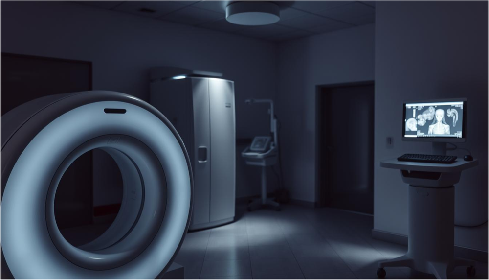
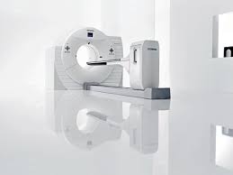
Computed Tomography (CT): Detailed Insights in Minutes

Computed Tomography (CT) scans give detailed images of the body, helping diagnose complex issues. CT scans are key in trauma cases to spot internal injuries fast. The clear images from CT scans help doctors make quick decisions.

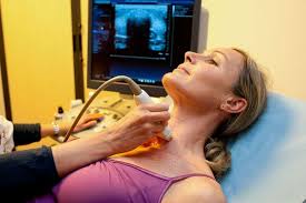
Ultrasound: Rapid Bedside Evaluation Capabilities

Ultrasound is great for quick bedside checks, helping make fast decisions. It’s used for checking abdominal pain, finding gallstones, and guiding procedures. Ultrasound’s portability and non-invasive nature make it perfect for emergencies.

MRI: When and Why It’s Used in Emergency Settings

MRI is not as common in emergencies because of its longer scan times and limited availability. But, it’s useful for diagnosing spinal cord injuries or certain brain conditions. MRI’s ability to see soft tissues makes it valuable in some emergency cases.

The choice of imaging tool in the emergency department depends on the situation, patient’s condition, and need for quick diagnosis. Knowing the strengths and weaknesses of each is key for the best patient care.

  • X-ray for initial assessments and fractures
  • CT for detailed insights and complex conditions
  • Ultrasound for rapid bedside evaluations
  • MRI for specific conditions requiring high soft tissue sensitivity

Trauma Imaging: When Seconds Count

When trauma happens, radiology plays a key role in saving lives. It does this by quickly diagnosing through imaging. The speed and accuracy of these results can mean the difference between life and death.

The Golden Hour: Optimizing Imaging Workflows in Trauma

The “golden hour” is a critical time in trauma care. It’s the first hour after an injury. Optimizing imaging workflows during this time is vital. It helps find injuries and start the right treatment fast.

Whole-Body CT Scanning in Multi-System Trauma

Whole-body CT scanning is a big help in multi-system trauma. It lets doctors check for injuries all over the body. This helps them focus on the most urgent treatments and can save lives.

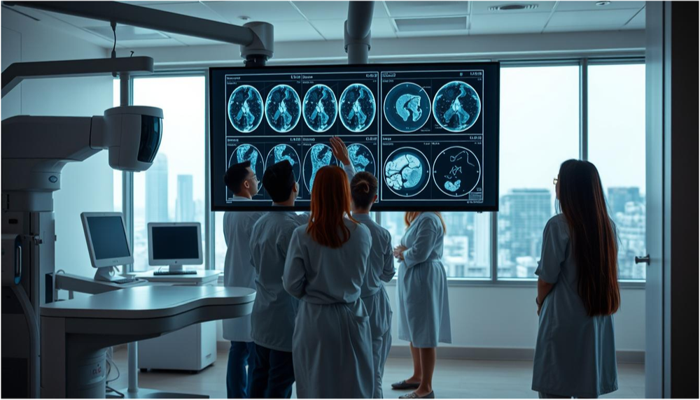
Detecting Life-Threatening Injuries: Brain, Chest, and Abdominal Trauma

Injuries to the brain, chest, and abdomen are very serious. Radiology is key in quickly spotting these injuries. This allows for quick action to save lives.

Case Study: How Rapid CT Saved a Multi-Trauma Patient

A multi-trauma patient was rushed to the emergency room. Quick CT scans helped doctors find and treat critical injuries fast. This saved the patient’s life.

In summary, trauma imaging is essential in emergency care. It helps diagnose and treat injuries quickly. This can greatly improve patient outcomes. Advanced imaging and efficient workflows are key to saving lives and helping patients recover.

Stroke Care Revolution: How Radiology Changed the Game

In emergency medicine, radiology is key in stroke care. The saying “time is brain” shows how fast we need to act in stroke cases.

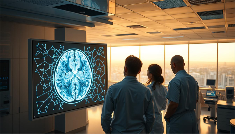
Time is Brain: The Critical Role of Rapid Imaging in Stroke

Rapid imaging is vital in stroke care. It lets doctors quickly see the stroke’s type and size. This info is key for choosing the right treatment.

CT Perfusion and Angiography in Acute Stroke Management

CT perfusion and angiography are now essential in managing acute strokes. They give detailed views of blood flow in the brain. This helps find the best places for treatment.

Extending the Treatment Window: How Advanced Imaging Makes It Possible

Advanced imaging has changed stroke care by giving more time for treatment. It lets doctors make better choices, even when time is running out.

Success Stories: Patients Saved by Timely Stroke Imaging

Many patients have been saved thanks to quick stroke imaging. These stories show how important fast and accurate imaging is in emergencies.

Chest Pain and Cardiac Emergencies: Imaging Decision Pathways

Effective imaging decision pathways are key for patients with chest pain and cardiac emergencies. It’s vital to quickly and accurately diagnose life-threatening conditions in emergency medicine.

Ruling Out Life-Threatening Conditions: Aortic Dissection and Pulmonary Embolism

When chest pain occurs, it’s important to rule out severe conditions like aortic dissection and pulmonary embolism. Imaging techniques are essential for diagnosing these conditions. For example, CT angiography is great for finding aortic dissections. A CT pulmonary angiogram is used to spot pulmonary embolisms.

Cardiac CT in the Emergency Setting

Cardiac CT is a valuable tool in emergencies. It gives detailed images of the heart and its vessels. It helps check for coronary artery disease, find cardiac anomalies, and look at the heart’s structure and function.

Radiation Dose Considerations in Cardiac Imaging

Cardiac imaging, like CT scans, is critical but we must think about radiation dose. It’s important to lower radiation exposure to prevent harm. Using dose modulation and the lowest necessary dose for images is becoming more common.

By improving imaging decision pathways and watching radiation exposure, healthcare providers can better care for patients in cardiac emergencies.

Pediatric Emergency Radiology: Special Considerations

Emergency radiology for kids is different because of their growing bodies and the need for safe radiation use.

Child-Specific Imaging Protocols and Radiation Safety

Imaging for kids needs special settings based on their size and age. Radiation safety is key. Protocols must reduce exposure while keeping images clear.

Non-Accidental Trauma: The Radiologist’s Role in Detection

Radiologists are vital in spotting non-accidental trauma in kids. Signs like different healing stages of fractures can show abuse.

Common Pediatric Emergencies and Their Imaging Findings

Common emergencies in kids include appendicitis, intussusception, and fractures. Knowing the usual imaging signs for these is essential for quick and correct diagnosis.

In summary, pediatric emergency radiology needs a careful approach. It must balance finding the right diagnosis with keeping radiation use safe.

The Expertise Behind Emergency Radiology: Training and Skills

Expertise in emergency radiology comes from deep education and ongoing learning. Emergency radiologists need specialized training to handle imaging in urgent situations.

Specialized Training for Emergency Radiologists

Emergency radiologists get a lot of education and training. This includes:

  • Completing a residency program in radiology
  • Participating in fellowship programs focused on emergency radiology
  • Engaging in continuous professional development to stay updated with the latest imaging techniques and technologies

The Canadian Perspective: Education and Certification

In Canada, radiologists must meet certain education and certification standards. This involves:

  1. Obtaining a medical degree from an accredited institution
  2. Completing a radiology residency program approved by the Royal College of Physicians and Surgeons of Canada
  3. Obtaining certification in radiology from the Royal College

Continuous Learning in a Rapidly Evolving Field

The field of emergency radiology keeps changing, with new tech and methods coming out all the time. To keep up, radiologists must keep learning, including:

  • Attending conferences and workshops focused on emergency radiology
  • Participating in online courses and webinars
  • Engaging in peer review and quality improvement initiatives

By combining initial training with ongoing education, emergency radiologists can offer top care in emergency medicine.

Technological Innovations Transforming Emergency Radiology

Technological advancements are changing emergency radiology for the better. They make patient care faster and more accurate. This leads to better health outcomes for patients.

Artificial Intelligence Applications in Emergency Imaging

Artificial intelligence (AI) is now a big part of emergency imaging. AI helps spot problems and sort out urgent cases. It also helps doctors make more precise diagnoses.

Point-of-Care Ultrasound: Bringing Imaging to the Bedside

Point-of-care ultrasound (POCUS) is key in emergency rooms. It lets doctors do ultrasound tests right at the patient’s side. This speeds up decision-making and improves care.

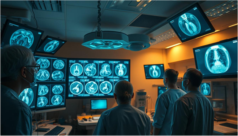
Advanced Visualization Tools for Faster Interpretation

Advanced tools are making it easier to understand radiology images. These tools improve image quality and give more detailed info.

3D Reconstruction Techniques

3D reconstruction techniques create detailed 3D images from radiology data. They’re very helpful in complex cases, like trauma or surgery planning.

Dual-Energy CT Applications

Dual-energy CT makes CT scans even better. It helps identify different tissues and materials, making diagnoses more accurate.

These new technologies are making emergency radiology better. They help doctors provide faster and more effective care. As technology keeps improving, we’ll see even more exciting advancements.

Overcoming Challenges in Modern Radiology for Emergency Care

Modern radiology in emergency settings faces many challenges. It must provide timely and effective care. Radiology is key in emergency medicine, but it struggles to deliver top-notch services.

Managing High Patient Volumes and Prioritization

Emergency departments see a lot of patients. It’s important to quickly sort out who needs help the most. Efficient triage systems help make sure urgent cases get attention fast.

  • Implementing advanced triage protocols
  • Utilizing AI for preliminary assessments
  • Streamlining workflows to reduce wait times

Radiation Dose Optimization Strategies

Reducing radiation doses is key to keeping patients safe. In emergencies, quick diagnosis is vital. Strategies include:

  1. Using low-dose protocols for common examinations
  2. Implementing dose management software
  3. Training staff on dose optimization techniques

Addressing Disparities in Access to Advanced Imaging

Not everyone has access to advanced imaging. This affects patient care. Efforts to fix this include:

  • Upgrading imaging equipment in underserved areas
  • Implementing teleradiology services
  • Promoting education and training for healthcare professionals

Canadian Healthcare System: Unique Challenges and Solutions

The Canadian healthcare system has its own challenges. One is unequal access to healthcare. Solutions include:

  • Standardizing emergency radiology protocols nationwide
  • Investing in telemedicine and remote imaging services
  • Enhancing training programs for radiologists and technicians

Conclusion: The Future of Emergency Radiology and Patient Care

The future of emergency radiology looks bright. New technologies will make imaging better, helping patients more. Radiology’s role in emergency care will grow, improving patient outcomes.

Artificial intelligence and advanced tools will change how radiology works. They will make diagnoses more accurate and care better. The Canadian healthcare system will face challenges, but these tools can help.

It’s key to keep training radiology professionals. They need to know how to use new tech. This way, emergency radiology can keep delivering top-notch care.

FAQ

What is the role of radiology in emergency medicine?

Radiology is key in emergency medicine. It helps diagnose quickly and saves lives. This is done through medical imaging technologies.

What are the most common imaging modalities used in emergency departments?

In emergency departments, common imaging tools include X-ray, CT, Ultrasound, and MRI.

How does radiology contribute to stroke care?

Radiology has changed stroke care a lot. It allows for fast imaging. This is vital for quick treatment, thanks to CT perfusion and angiography.

What are the unique challenges faced by radiologists in emergency settings?

Radiologists in emergencies face big challenges. They must be fast but also accurate. They need special training and skills for high-pressure situations.

How is pediatric emergency radiology different from adult emergency radiology?

Pediatric emergency radiology is different. It focuses on child safety and uses special imaging. It also looks for signs of abuse.

What technological innovations are transforming emergency radiology?

New tech is changing emergency radiology. This includes AI, ultrasound, advanced tools, 3D images, and dual-energy CT.

How is radiation dose optimized in emergency imaging?

Minimizing radiation risk is key in emergency imaging. This is done with child-safe protocols and choosing the right imaging.

What is the significance of the “golden hour” in trauma imaging?

The “golden hour” is very important in trauma care. It highlights the need for fast imaging. Quick diagnosis and treatment can save lives.


Federated Learning in Medical Imaging: Privacy-Preserving AI Across Hospitals

Federated Learning is changing the game in Medical Imaging. It lets hospitals work together on AI projects without sharing patient data. This keeps patient privacy safe.

This new method helps healthcare places share knowledge and insights. They do this without giving out personal patient data. This speeds up the growth of AI in healthcare.

With Federated Learning, hospitals can make better diagnoses and treatments. They do this while keeping patient info private.

This introduction prepares us for a closer look at how Federated Learning is changing Medical Imaging and AI in healthcare.

The Data Privacy Challenge in Healthcare AI

Healthcare AI systems struggle to balance data needs with patient privacy. The use of artificial intelligence in medical care has made data privacy a big concern. It’s a key issue in the healthcare world.

Patient Data Protection Regulations

The healthcare industry must follow strict rules to protect patient data. Two important rules stand out:

  • HIPAA and Canadian Privacy Laws: In the U.S., HIPAA protects patient information. Canada has its own privacy laws for healthcare providers.
  • Cross-border Data Sharing Limitations: Sharing patient data across borders is tricky. Different countries have different data protection rules.

Traditional AI Training Limitations

Traditional AI training methods use big datasets in one place. This is a big privacy risk. Moving sensitive patient data to a central server for training can lead to breaches.

The Need for Collaborative Learning Solutions

Traditional AI training has its limits. We need new ways to train AI that keep patient data safe. Federated Learning is a good solution. It trains AI models on data that stays local, keeping patient info private.

  1. Federated Learning trains AI models on data that stays local. This reduces the risk of data breaches.
  2. This method helps healthcare institutions work together without risking patient privacy.

Understanding Federated Learning: A Primer

Federated Learning is key for privacy in AI, vital in healthcare, like in medical imaging in hospitals.

Definition and Core Concepts

Federated Learning lets many sites work together on model training without sharing data. Models are trained locally on each site’s data. Then, only the model updates are shared, not the data itself.

The main idea is decentralized data processing. This keeps sensitive information safe at each site.

How Federated Learning Differs from Centralized AI

Federated Learning and traditional AI differ in data processing. Centralized AI gathers data in one place for training.

But Federated Learning trains models locally at each site. This keeps data private.

Decentralized Model Training

This method trains models on various data sets without moving data. It keeps privacy safe.

Local Computation vs. Data Sharing

By doing computations locally and sharing updates, Federated Learning lowers data sharing risks. This includes privacy breaches.

The Technical Architecture of Federated Systems

Federated Learning systems have a central server for coordinating model training. Different nodes, like hospitals, train local models.

Updates from each node are sent to the server. There, they’re combined to create a strong AI model.

The Revolution in Medical Imaging Through Federated Learning

Federated Learning is changing medical imaging by making AI development collaborative and private. It lets many healthcare places work together to train AI models on different data sets.

Federated Learning in Medical Imaging: Privacy-Preserving AI Across Hospitals

Federated Learning in Medical Imaging: Privacy-Preserving AI Across Hospitals

Evolution of AI in Diagnostic Imaging

AI in medical imaging has made diagnoses more accurate. Federated Learning makes these models even better by training them together across different places.

Privacy-Preserving Model Training

Federated Learning trains AI models on sensitive medical images without sharing the data. It does this by sharing updates of the model, keeping patient information safe.

Improving Diagnostic Accuracy Across Diverse Populations

Federated Learning uses data from many places to improve diagnosis. This is great for spotting rare or varied conditions in different groups.

Reducing Bias in Image Analysis

Federated Learning reduces bias in AI by training on a wide variety of images. This makes healthcare fairer by working well for all kinds of patients.

Enhancing Rare Condition Detection

Federated Learning helps find rare conditions by combining data from many places. This teamwork boosts the ability to diagnose uncommon diseases.

In summary, Federated Learning is set to change medical imaging. It will make diagnoses better, reduce bias, and find rare conditions, all while keeping patient info private.

Federated Learning Applications in Radiology

Federated learning is changing radiology by letting hospitals work together on AI training. They do this without sharing patient data. This teamwork is making diagnoses better in many imaging types.

Chest X-ray Analysis Advancements

Federated learning has made big strides in chest X-ray analysis. AI models now spot problems like pneumonia and heart issues more accurately. This is thanks to using data from many hospitals.

Key advancements include:

  • Enhanced detection accuracy
  • Improved model generalizability across different patient populations
  • Reduced need for centralized data storage, boosting privacy

CT Scan Interpretation

Federated learning is also changing CT scan analysis. It helps create smarter AI models. These models can spot complex issues and help find diseases early.

MRI Processing Techniques

In MRI processing, federated learning is making big leaps. It’s improving how we analyze images, like brain and muscle scans.

Brain Imaging Applications

Federated learning is helping create AI for brain tumor detection and neurodegenerative disease spotting. It uses MRI scans for this.

Musculoskeletal Diagnostics

In musculoskeletal diagnostics, federated learning is making MRI scans better. It helps find issues like osteoarthritis and muscle injuries more clearly.

Implementation Challenges for Hospital Networks

Federated learning is promising but comes with big challenges for hospital networks. These include technical issues and managing data. Healthcare institutions must tackle these to make federated learning work well.

Technical Infrastructure Requirements

The tech needed for federated learning in hospitals is a lot. It’s not just about computers and networks. It also means working with current systems for storing and sharing medical images.

Computing Resources and Network Capabilities

Hospitals need strong computers and networks for federated learning. This means:

  • High-performance computing nodes for big data
  • Advanced network infrastructure for quick data sharing
  • Robust security measures to keep patient data safe

Integration with Existing PACS Systems

Working with current PACS systems is key for federated learning. This means:

  1. Creating custom APIs for smooth integration
  2. Keeping data consistent across systems
  3. Ensuring high data integrity during sharing

Data Heterogeneity Issues

Data from different hospitals can be a big problem for federated learning. Each place might have different data types, quality, and how it’s collected. This makes training models harder.

Computational Resource Management

Managing computer resources well is key for federated learning. It’s about:

  • Allocating resources wisely
  • Managing how work is spread out
  • Scaling the system as needed

By solving these issues, hospitals can use federated learning to better diagnose and treat patients in radiology.

Privacy Preservation Mechanisms in Healthcare Federated Systems

Protecting sensitive medical information is key in healthcare federated systems. Federated Learning lets different healthcare places work together on AI models. They do this without sharing raw patient data, which helps keep things private.

Differential Privacy Techniques

Differential privacy adds noise to data or model updates. This makes it hard to identify individual patient info. It’s very useful in healthcare federated systems where data is spread out.

Key benefits include:

  • Enhanced patient privacy
  • Compliance with healthcare regulations
  • Robustness against data breaches

Secure Aggregation Protocols

Secure aggregation protocols let different healthcare providers add their model updates together. They do this without showing what each update is. This is done with special cryptography that keeps each hospital’s data private.

Homomorphic Encryption in Clinical Settings

Homomorphic encryption lets you do computations on encrypted data. It’s perfect for keeping AI in healthcare private. In clinics, it means patient data stays encrypted while training AI models.

Practical Applications in Radiology Departments

Radiology departments can really benefit from homomorphic encryption. They can analyze encrypted medical images. This helps make AI models for disease diagnosis more accurate without risking patient privacy.

Performance Considerations

Homomorphic encryption is great for privacy but can slow things down. Making these algorithms faster is important for using them in healthcare. This is true for many hospitals working together.

Using these privacy tools, healthcare federated systems can keep patient data safe. They can also improve AI in medical imaging and diagnostics.

Case Studies: Successful Federated Learning Across Hospitals

Hospitals can now work together on AI model training safely. This method has greatly improved medical imaging diagnostics.

Multi-institutional Chest X-ray Analysis Networks

Hospitals have joined forces to analyze chest X-rays using federated learning. This teamwork has made spotting problems better and improved how well doctors can diagnose.

Key benefits include:

  • Improved model robustness
  • Enhanced patient care
  • Better handling of diverse data sets

Federated Brain MRI Tumor Detection Projects

Federated learning has been a hit in finding brain MRI tumors. By combining data from different places, scientists have made more precise tumor models.

Cross-border Collaboration in Rare Disease Imaging

Working together across borders has helped build big datasets for rare diseases. This is super helpful for diseases where there’s little data.

Canadian-US Hospital Partnerships

Canadian and US hospitals have teamed up to make strong AI models for rare disease diagnosis. These partnerships have shown great promise in boosting diagnostic accuracy.

Measurable Outcome Improvements

The results of these partnerships are clear, with big wins in diagnostic accuracy and patient care. The success of these projects shows how powerful federated learning can be in medical imaging.

The Future of Medical Imaging with Federated Learning

Medical imaging is on the verge of a new era thanks to federated learning. This method of AI development lets different places work together on models. It keeps data safe and private.

Emerging Research Directions

Research in federated learning for medical imaging is growing fast. It’s looking into better algorithms for diverse data, more accurate models, and less need for data labeling.

Key areas of focus include making models easier to understand, dealing with different data types, and finding new uses in various imaging types.

Integration with Other Privacy-Preserving Technologies

Federated learning is being paired with other tech to boost data safety. Two key examples are:

  • Blockchain for Model Verification: Blockchain helps keep a record of model changes. It makes sure everyone trusts the learning process.
  • Zero-Knowledge Proofs in Healthcare: Zero-knowledge proofs let models be checked without sharing personal data. This adds more privacy.

Potential for Global Healthcare Collaboration

Federated learning could lead to big changes in global healthcare. It lets hospitals and research centers worldwide work together on AI. This could make models better and more accurate for different people.

This teamwork could bring big advances in medical imaging. It could lead to new ideas and better healthcare for everyone.

Conclusion: Balancing Innovation and Privacy in Healthcare AI

Federated Learning is changing Healthcare AI. It lets hospitals work together on AI models while keeping patient data safe. This is very important in radiology, where it helps make diagnoses more accurate.

Healthcare places can use Federated Learning to innovate in medical imaging safely. This way, AI models get better at diagnosing diseases. Patients then get better care.

The future of Healthcare AI looks bright with technologies like Federated Learning. They focus on both new ideas and keeping data safe. We’ll see big improvements in radiology and other imaging areas.

FAQ

What is Federated Learning, and how does it apply to medical imaging?

Federated Learning is a way for different groups to work together on training models without sharing data. In medical imaging, it lets hospitals and research centers train AI models together. They do this using their own data, keeping patient information safe.

How does Federated Learning address data privacy concerns in healthcare AI?

Federated Learning keeps patient data safe by not sharing it. Only model updates are shared, which are small and less risky. This meets privacy laws, making it easier to work on AI together.

What are the benefits of using Federated Learning in radiology?

Federated Learning in radiology boosts accuracy and reduces bias. It helps spot rare conditions better. It also makes AI models stronger, working well with different images and scans.

What technical infrastructure is required for implementing Federated Learning in hospital networks?

To use Federated Learning, hospitals need strong tech. This includes good computers, fast networks, and systems for sharing updates. They also need experts to keep everything running smoothly and securely.

How does Federated Learning handle data heterogeneity across different hospitals?

Federated Learning uses special techniques to handle different data. It normalizes and adapts data to make models work well everywhere. This improves how well models can diagnose diseases.

What are some of the emerging research directions in Federated Learning for medical imaging?

New research is looking at combining Federated Learning with other tech for more security. It’s also exploring new uses, like finding rare diseases and personalized medicine.

Can Federated Learning be used for global healthcare collaboration?

Yes, Federated Learning can help countries work together on AI. It lets them train models on their own data. This creates stronger models that can help patients all over the world.

AI-Enhanced Multi-Modality Imaging: Combining CT, MRI, and Ultrasound for Smarter Diagnoses

Medical diagnosis has seen a big change with AI-enhanced imaging. Now, doctors can use CT, MRI, and ultrasound together. This helps them make more accurate diagnoses.

By mixing these technologies with AI algorithms, doctors can spot complex patterns. This leads to smarter diagnoses. It also gives a better understanding of what’s going on with the patient.

So, doctors can create better treatment plans. This improves how well patients do. The mix of AI Imaging and medical knowledge is changing diagnostics.

The Current Landscape of Medical Imaging

Medical imaging has made big strides, but it’s not perfect yet. There are issues with how accurate it is and what it can do. Radiology is always getting better, aiming for more precise and reliable ways to diagnose.

Challenges in Traditional Diagnostic Imaging

Methods like CT scans, MRI, and ultrasound are key in medical diagnosis. But, they have their own problems.

Limitations of Single-Modality Approaches

Using just one imaging method can miss the mark. For example, ultrasound depends a lot on the person doing it. And CT scans might not show soft tissues well enough.

Diagnostic Accuracy Concerns

Getting a correct diagnosis is a big deal in medical imaging. Single-method approaches can lead to wrong or late diagnoses. This can really affect how well patients do.

The Need for Integrated Approaches

Using more than one imaging method can give a fuller picture of what’s going on inside the body.

Complementary Information from Multiple Sources

Putting together data from CT, MRI, and ultrasound can help doctors feel more sure and accurate in their diagnoses.

Evolution Toward Precision Medicine

This way of combining imaging is moving us toward precision medicine. It means treatments will be more tailored to each patient, based on detailed diagnostic info.

The move to using more imaging methods and combining them is changing radiology. It’s helping doctors make better choices and improving care for patients.

Understanding Individual Imaging Modalities


Advanced medical imaging techniques like CT, MRI, and ultrasound have changed how we diagnose diseases. Each method has its own way of working, benefits, and drawbacks.

Computed Tomography (CT): Principles and Limitations

CT scans use X-rays to make detailed images of the body’s inside. CT scans are very useful in emergency situations, like finding internal injuries or bleeding.

Radiation Exposure Considerations

One big problem with CT scans is the ionizing radiation they use. This can raise the risk of cancer. But, the benefits often outweigh the risks, mainly in urgent care.

Optimal Applications in Diagnosis

CT scans are best for finding tumors, fractures, and vascular diseases. They give quick and accurate results, which are key in emergency medicine.

Magnetic Resonance Imaging (MRI): Capabilities and Constraints

MRI uses strong magnetic fields and radio waves to make detailed images of the body’s inside. MRI is great at showing soft tissues, which is why it’s so valuable for diagnosing brain, spinal cord, and joint problems.

Soft Tissue Visualization Advantages

MRI’s ability to show soft tissues in detail is a big plus. It helps diagnose things like ligament tears, herniated discs, and some cancers.

Time and Cost Factors

One drawback of MRI is how long it takes. It’s also more expensive than CT scans or ultrasound.

Ultrasound Technology: Strengths and Weaknesses

Ultrasound uses sound waves to create images of the body’s inside. It’s a safe and non-invasive method that’s used a lot for diagnosis.

Real-Time Imaging Benefits

Ultrasound’s biggest advantage is its ability to show images in real-time. This is really helpful during things like biopsies or watching a fetus grow.

Operator Dependency Challenges

But, ultrasound depends on the person doing the scan. The quality of the images can vary a lot based on the operator’s skill.

The Rise of AI Imaging in Modern Medicine

Artificial Intelligence is now a big part of radiology. It helps improve how we analyze and understand medical images. This change is making diagnosis faster and more accurate.

AI-Enhanced Multi-Modality Imaging: Combining CT, MRI, and Ultrasound for Smarter Diagnoses

AI-Enhanced Multi-Modality Imaging: Combining CT, MRI, and Ultrasound for Smarter Diagnoses

Machine Learning Algorithms in Image Analysis

Machine learning algorithms are key in medical image analysis. They come in two types: supervised and unsupervised learning.

Supervised Learning for Classification Tasks

Supervised learning uses labeled data to teach algorithms. It’s great for spotting specific health issues in images.

Unsupervised Learning for Pattern Recognition

Unsupervised learning finds patterns in data without labels. It uncovers new insights and oddities in medical images.

Deep Learning Applications in Radiology

Deep learning, like Convolutional Neural Networks (CNNs), is making waves in radiology.

Convolutional Neural Networks (CNNs)

CNNs excel at recognizing patterns in images. This skill is vital for image classification tasks.

Transfer Learning Approaches

Transfer learning refines pre-trained models for radiology. It boosts performance and cuts down on the need for lots of labeled data.

Computer Vision Techniques for Medical Images

Computer vision is being used more in medical imaging. It’s for tasks like segmenting images, extracting features, and finding anomalies.

Segmentation and Feature Extraction

Segmentation isolates specific parts of medical images. Feature extraction measures these parts’ characteristics.

Anomaly Detection Methods

Anomaly detection spots unusual things in images. It’s key for early diagnosis and planning treatments.

Technique Application Benefit
Supervised Learning Classification Tasks High Accuracy
Unsupervised Learning Pattern Recognition Discovers New Insights
CNNs Image Classification Complex Pattern Recognition

How AI Enhances Each Imaging Modality

Artificial intelligence is changing medical imaging. It’s making CT, MRI, and ultrasound better. AI algorithms are improving image quality and analysis.

AI-Powered CT Image Reconstruction and Analysis

AI is transforming CT imaging. It’s improving how images are made and analyzed. Noise reduction and image quality improvement are key areas where AI shines.

Noise Reduction and Image Quality Improvement

AI is making CT images clearer. It reduces noise, helping doctors spot small problems. This is vital for accurate diagnoses.

Automated Lesion Detection

AI can find lesions in CT scans automatically. This helps doctors spot issues fast and accurately.

MRI Enhancement Through Artificial Intelligence

AI is boosting MRI technology. It’s improving accelerated acquisition techniques and quantitative analysis capabilities.

Accelerated Acquisition Techniques

AI is making MRI scans faster. This makes them more comfortable for patients and more efficient for machines.

Quantitative Analysis Capabilities

AI also allows for detailed MRI analysis. It gives insights into tissue and pathology.

Ultrasound Interpretation with AI Assistance

In ultrasound, AI helps with real-time guidance systems and automated measurements and calculations.

Real-Time Guidance Systems

AI-assisted ultrasound systems offer real-time feedback. This boosts procedure accuracy.

Automated Measurements and Calculations

AI also automates ultrasound tasks. This saves healthcare professionals time and reduces errors.

Multi-Modality Integration: Technical Foundations

Multi-modality imaging combines different techniques like CT, MRI, and ultrasound. This gives a full view of what’s going on inside the body. It’s all thanks to advanced tech that lets data from various sources come together.

Data Fusion Techniques for Medical Images

Data fusion is key for mixing info from different imaging types. Early fusion and late fusion are the main methods used in medical imaging.

Early Fusion vs. Late Fusion Approaches

Early fusion blends data at the image level. Late fusion, on the other hand, combines the results of each modality’s analysis. The choice depends on the clinical need.

Multiparametric Analysis Methods

Multiparametric analysis looks at many features from different imaging types. This method boosts accuracy by giving a detailed look at a patient’s health.

Co-registration and Alignment Challenges

Aligning images from different sources is vital for accurate fusion. Rigid and non-rigid registration algorithms help match images.

Rigid and Non-rigid Registration Algorithms

Rigid registration works for images that have only moved slightly. Non-rigid registration is for images that have changed shape.

Temporal Alignment Considerations

Temporal alignment is key for dynamic imaging. It makes sure images from different times match up.

Creating Comprehensive 3D Visualizations

Advanced visualization creates detailed 3D views of the combined data. Volume rendering techniques and interactive visualization tools help doctors understand complex images better.

Volume Rendering Techniques

Volume rendering makes 3D images from 3D data. It shows what’s inside the body.

Interactive Visualization Tools

Interactive tools let doctors play with 3D images. They can look at data from all sides, helping with diagnosis.

Clinical Applications of AI-Enhanced Multi-Modality Imaging

AI-enhanced multi-modality imaging has many uses in medicine. It helps in several key areas of medical practice. This technology is used in many medical fields to improve diagnosis and treatment.

Oncology: Tumor Detection and Characterization

In oncology, AI plays a big role in finding and understanding tumors. By mixing data from CT, MRI, and ultrasound, AI gives more accurate diagnoses.

Improved Staging and Treatment Planning

AI imaging helps in precise tumor staging. This is key for choosing the best treatment. For example, CT and PET data together show how far tumors have spread.

Monitoring Treatment Response

AI can track changes in tumors over time. This lets doctors see if treatments are working early on.

Neurology: Brain Mapping and Disorder Diagnosis

In neurology, AI helps map the brain and diagnose disorders. MRI is very useful here.

Stroke Assessment and Management

AI imaging quickly checks how severe a stroke is. It also finds the best treatment.

Neurodegenerative Disease Evaluation

Cardiovascular Applications: Structural and Functional Assessment

In heart medicine, AI imaging checks the heart’s structure and function.

Coronary Artery Disease Evaluation

CT angiography with AI checks coronary artery disease.

Cardiac Function Analysis

AI analyzes ultrasound and MRI data to check heart function.

Musculoskeletal Imaging: Improving Precision and Accuracy

In musculoskeletal imaging, AI makes diagnoses more precise and accurate.

Joint and Soft Tissue Assessment

MRI and ultrasound together check joints and soft tissues.

Trauma and Degenerative Disease Evaluation

AI imaging helps evaluate trauma and degenerative diseases.

Medical Specialty Primary Imaging Modalities AI-Enhanced Applications
Oncology CT, MRI, Ultrasound Tumor detection, staging, treatment response monitoring
Neurology MRI Brain mapping, stroke assessment, neurodegenerative disease evaluation
Cardiovascular CT, Ultrasound, MRI Coronary artery disease evaluation, cardiac function analysis
Musculoskeletal MRI, Ultrasound Joint and soft tissue assessment, trauma and degenerative disease evaluation

Benefits and Outcomes of Integrated Imaging Approaches

AI-powered imaging is changing how we diagnose and treat patients. It combines CT, MRI, and ultrasound to give doctors a clearer picture. This leads to more accurate and detailed diagnoses.

Improved Diagnostic Accuracy and Confidence

AI makes diagnoses better by analyzing complex data. AI algorithms can spot patterns that humans might miss.

Reduction in False Positives and Negatives

AI helps avoid wrong diagnoses. This is key in radiology, where getting it right is critical.

Enhanced Lesion Characterization

AI can better understand lesions. It gives detailed info on their nature and size. This helps doctors plan treatments.

Reduced Need for Invasive Procedures

AI reduces the need for risky tests. Non-invasive tissue analysis makes exams safer and more comfortable for patients.

Non-invasive Tissue Characterization

AI supports detailed tissue analysis without biopsies. This is a big step forward in diagnostics.

Virtual Biopsy Concepts

AI is making virtual biopsies possible. This could change how we diagnose diseases, making it safer and more precise.

Time and Cost Efficiency in the Diagnostic Process

AI speeds up and saves money in diagnosis. It makes the process more efficient and cost-effective.

Streamlined Workflow Integration

AI fits into current workflows, boosting efficiency. This helps doctors work better and faster.

Resource Optimization Strategies

AI helps use resources wisely. This cuts costs and improves patient care.

Patient-Centered Advantages

AI imaging benefits patients in many ways. It offers personalized care and better experiences.

Personalized Diagnostic Approaches

AI tailors care to each patient. This improves the quality of care for everyone.

Improved Patient Experience

AI reduces the need for risky tests. This makes the diagnostic process smoother and safer for patients.

Implementation Challenges and Considerations

Using AI in multi-modality imaging, like CT and ultrasound, is not easy. It needs a big effort to get past technical, regulatory, and educational barriers.

Technical Infrastructure Requirements

To support AI in imaging, you need a lot of tech. This includes fast computers and lots of storage for big data.

Computing Resources and Storage Needs

Advanced tech is key for handling complex data. High-capacity storage solutions are also needed to store all the data.

Network and Integration Considerations

Connecting different imaging types and AI needs a strong network. It’s important for systems to work well together.

Regulatory and Approval Pathways

Getting AI imaging approved is a big challenge. You must follow rules to use it in clinics.

FDA and Health Canada Clearance Processes

Knowing the rules from the FDA and Health Canada is important. These rules make sure AI imaging is safe and works well.

Validation Requirements for Clinical Use

AI and imaging systems must be tested well. This shows they are useful and accurate in clinics.

Training Requirements for Healthcare Professionals

Healthcare workers need good training for AI imaging. They must learn how to use and understand AI insights.

Radiologist Education and Adaptation

Radiologists and others need to learn about AI tools. They must know what AI can do and what it can’t.

Multidisciplinary Team Collaboration

Working together is key for AI imaging success. Teams of experts are needed to make the most of AI.

Ethical and Privacy Considerations

AI in imaging raises big questions about ethics and privacy. Keeping patient data safe is very important.

Data Security and Patient Confidentiality

Protecting patient info is a must. Following privacy rules is essential.

Algorithmic Transparency and Bias Mitigation

AI tools must be clear and fair. Regular checks and updates help keep trust in AI.

AI is changing how we do medical imaging, like MRI and other radiology methods. By using CT, MRI, and ultrasound together, doctors can understand patients better. This leads to more accurate diagnoses and treatments.

AI algorithms are getting better, making radiology more precise. This means doctors can analyze complex images with great accuracy. The future of AI in imaging looks very promising for better patient care and easier clinical work.

To make the most of AI in imaging, we need to keep investing in technology, rules, and training doctors. With AI, the medical field is set for a new era of excellence in diagnosis and care.

FAQ

What is AI-enhanced multi-modality imaging?

AI-enhanced multi-modality imaging uses different medical imaging tech like CT, MRI, and ultrasound. It adds artificial intelligence to boost how well doctors can diagnose and feel sure about their findings.

How does AI enhance medical imaging modalities like CT, MRI, and ultrasound?

AI makes these imaging methods better by improving how images are made, analyzed, and understood. For instance, AI can clean up CT images, speed up MRI scans, and guide ultrasound procedures in real-time.

What are the benefits of using AI-enhanced multi-modality imaging in clinical practice?

Using AI in imaging helps doctors make more accurate diagnoses. It also means fewer invasive tests, better patient experiences, and a smoother workflow. All these lead to better health outcomes for patients.

What are the challenges in implementing AI-enhanced multi-modality imaging?

There are a few hurdles to overcome. These include setting up the right tech, getting approvals, training staff, and thinking about ethics like keeping patient data safe and being open about how AI works.

How does AI-enhanced multi-modality imaging impact patient care in oncology?

In cancer care, AI helps spot tumors better and understand their size and type. It also helps plan treatments and check how well they’re working. This leads to more tailored and effective cancer care.

What role does radiology play in AI-enhanced multi-modality imaging?

Radiology is key in using AI for imaging. Radiologists look at images and work with AI to make diagnoses more accurate and confident.

Are there any specific regulations or guidelines for the use of AI in medical imaging in Canada?

Yes, Health Canada and other groups have rules and checks for AI in imaging. These ensure it’s safe and works well.

How does AI-enhanced multi-modality imaging contribute to precision medicine?

AI imaging helps precision medicine by giving detailed, personal info for diagnosis. This allows for targeted treatments and better health results for patients.


Reducing Diagnostic Bias: How AI Algorithms Are Shaping Equitable Radiology

Equitable healthcare is a basic right, but diagnostic bias is a big challenge. Artificial intelligence (AI) is becoming key in solving this problem.

AI is changing radiology by making diagnoses more accurate and fair. It looks at lots of data to find patterns that doctors might miss. This leads to better care for all patients.

AI in healthcare is more than just new tech. It’s about making sure every patient gets a diagnosis that fits their health, not just based on biases.

The Persistent Challenge of Bias in Diagnostic Imaging

Diagnostic imaging is key in healthcare but faces biases that harm patient care. These biases can cause unfair differences in who gets imaging services and how accurate they are. This affects the quality of care everyone receives.

Understanding Disparities in Medical Imaging Access

Some groups face big challenges getting medical imaging. This is due to things like money and where they live. These issues make it hard for some to get the care they need.

  • Socioeconomic status affecting access to quality imaging
  • Geographic barriers limiting availability of diagnostic services
  • Lack of diversity in clinical trials for imaging technologies

Demographic Factors Influencing Diagnostic Accuracy

Things like age, gender, and ethnicity can change how well imaging works. For example, some imaging might not work as well for people of different body types.

Key demographic factors include:

  1. Age-related changes affecting image interpretation
  2. Gender differences in disease presentation
  3. Ethnic variations in anatomical characteristics

Economic and Geographic Barriers to Quality Healthcare

Money and location can block access to good healthcare, including imaging. It’s important to tackle these issues to make healthcare fair for all.

Modern Radiology: Where Human Judgment Meets Technological Limitations

Reducing Diagnostic Bias: How AI Algorithms Are Shaping Equitable Radiology

Reducing Diagnostic Bias: How AI Algorithms Are Shaping Equitable Radiology

The field of radiology is at a crossroads. Here, human judgment and technology meet, shaping how we diagnose. Even with new imaging tech, human errors and tech limits affect how accurate our diagnoses are.

Cognitive Biases Among Imaging Specialists

Imaging specialists, like all humans, face cognitive biases. Biases like confirmation and anchoring bias can lead to misreading or missing important details in images.

Dataset Limitations and Historical Inequities

The data used to train AI in radiology often shows old biases. This means AI might not work well for everyone, making healthcare unfair.

Training Gaps and Their Consequences for Patient Outcomes

Training gaps in radiologists and technologists can harm patients. Not learning new tech or methods can cause mistakes in reading images and diagnosing.

To fix these issues, we need a plan. This plan should include better education, new tech, and fairness in radiology.

The Evolution of Artificial Intelligence in Medical Imaging

The use of artificial intelligence (AI) in medical imaging has changed how we diagnose diseases. AI has moved from simple systems to complex deep learning algorithms. This has made medical image analysis more accurate and efficient.

From Rule-Based Systems to Deep Learning

The start of AI in medical imaging was with rule-based systems. These systems followed set rules to understand images. But, they struggled with complex cases.

The introduction of deep learning changed everything. It allowed AI to learn from huge datasets. This made AI’s ability to diagnose better over time.

Key Technological Breakthroughs in Medical Image Analysis

Several key advancements have moved medical image analysis forward. The creation of convolutional neural networks (CNNs) has greatly improved image recognition. Also, computer vision advancements have helped in detecting and characterizing lesions.

This has helped radiologists make more precise diagnoses.

The Current Landscape of AI Tools in Clinical Practice

AI tools are now a big part of clinical practice. They help radiologists with tasks like image segmentation and disease detection. AI is making workflows smoother and improving patient care.

The field is seeing more AI solutions. Research is ongoing to make these tools even better and more useful in clinics.

How AI Algorithms Reduce Diagnostic Bias in Radiology

AI is changing radiology by cutting down on bias in diagnosis. It uses advanced algorithms to look at medical images with great detail. This is key to making sure diagnoses are fair and not swayed by personal opinions.

Pattern Recognition Beyond Human Capability

AI can spot patterns in medical images that humans can’t. It finds small changes in tissue or hidden issues. This skill is important for reducing bias, as it uses facts, not guesses.

Standardization of Diagnostic Criteria Across Populations

AI helps set the same rules for diagnosing in all patients. This makes sure everyone gets the same care. It’s a big step towards fair healthcare for all.

Key benefits of AI in standardizing diagnostic criteria include:

  • Consistency in diagnosis across different patient demographics
  • Reduced variability in diagnostic interpretations
  • Enhanced accuracy in detecting conditions

Removing Subjective Elements from Image Interpretation

AI uses numbers to understand images, not feelings. This cuts down on bias in diagnosis.

Quantitative Analysis of Subtle Imaging Features

AI looks at small details in images that people might miss. It gives a deeper look at what’s in medical images. This is important for making sure diagnoses are based on all the facts.

Consistent Application of Diagnostic Standards

AI always uses the same rules for diagnosing, no matter who the patient is. This makes healthcare fair for everyone.

Using AI in radiology helps doctors make fairer and more accurate diagnoses. This is a big step towards making healthcare fair for everyone.

Cutting-Edge AI Technologies Transforming Diagnostic Accuracy

AI is changing how we diagnose diseases in radiology. It’s making diagnoses more accurate and improving patient care.

Convolutional Neural Networks for Image Analysis

Convolutional Neural Networks (CNNs) are key in medical image analysis. They learn from images to spot patterns humans might miss. This is great for finding small issues in scans.

Computer Vision Advancements in Lesion Detection

AI in computer vision is making lesion detection better. It can look through lots of images fast and find problems early.

Natural Language Processing for Standardized Reporting

Natural Language Processing (NLP) helps make radiology reports the same. This makes it easier for doctors to talk about patient care, leading to better health outcomes.

Automated Report Generation and Quality Control

AI NLP systems can write radiology reports automatically. This saves time and reduces mistakes. It also makes reports more consistent.

Contextual Understanding of Medical Terminology

NLP lets AI systems get the meaning behind medical words. This makes reports clear and right, helping doctors work better together.

AI is changing radiology for the better. It’s making healthcare fairer and more effective. As AI gets smarter, it will help patients even more.

Case Studies: AI Success Stories in Promoting Healthcare Equity

Recent studies show AI’s success in making healthcare fairer for all. It uses artificial intelligence in radiology to cut down on bias and boost patient care.

Breast Cancer Detection Across Diverse Populations

AI has been a game-changer in spotting breast cancer in mammograms for all ethnic groups. A study found AI systems cut down on missed diagnoses by 30% in dense breasts, a big problem for young women.

Pulmonary Disease Identification in Underserved Communities

In areas where healthcare is scarce, AI helps spot lung diseases like TB better. AI checks chest X-rays and finds TB as well as doctors do.

Neurological Disorder Diagnosis Across Age and Ethnic Groups

AI helps diagnose brain diseases like Alzheimer’s and strokes for people of all ages and backgrounds. It looks at MRI and CT scans to spot early signs of these conditions.

Cardiac Imaging Interpretation in Global Settings

AI has made diagnosing heart problems worldwide better. It looks at heart scans to find issues like blocked arteries and heart failure.

Condition AI Application Outcome
Breast Cancer Mammography Analysis 30% reduction in false negatives
Pulmonary Disease Chest X-ray Analysis Improved detection rates in underserved communities
Neurological Disorders MRI and CT Scan Analysis Early detection of conditions like Alzheimer’s
Cardiac Conditions Echocardiogram Analysis Improved diagnosis of heart disease

These examples show AI’s power in radiology to make healthcare fairer. It improves accuracy for everyone, no matter where they are or who they are.

Challenges and Limitations in AI-Driven Diagnostic Systems

AI is becoming more important in radiology, but it faces many challenges. These systems have many benefits. Yet, they also have big hurdles to overcome before they can be safely used in hospitals.

Algorithm Transparency and the “Black Box” Problem

One big worry is how AI systems make decisions. This is called the “black box” problem. It’s hard for doctors to trust these systems because they don’t understand how they work.

Data Quality and Representation Issues

The data used to train AI is very important. But, this data is often biased or not diverse enough. This can lead to wrong or unfair results.

Regulatory and Ethical Considerations

There are also big issues with rules and ethics. It’s important to make sure AI systems follow all the rules and act ethically.

FDA Approval Processes for AI Medical Devices

The FDA has clear rules for approving AI medical devices. These rules help make sure AI systems are safe and work well.

Patient Privacy and Data Security Concerns

Keeping patient information safe is a big concern. It’s very important that AI systems protect this sensitive data.

Challenge Description Potential Solution
Algorithm Transparency Lack of understanding in AI decision-making Development of explainable AI models
Data Quality Biases and limitations in training data Diverse and representative datasets
Regulatory Compliance Ensuring adherence to regulatory standards Regular audits and compliance training

It’s very important to tackle these challenges for AI to work well in radiology. By fixing these issues, we can use AI to its fullest in medical imaging.

Implementing AI Solutions in Radiology Departments

AI is changing radiology fast. It’s key to use it well. Radiology teams need to smoothly add AI to their work.

Integration with Existing PACS and RIS Workflows

One big challenge is linking AI with PACS and RIS. Seamless integration helps keep work flowing. It makes sure AI insights are easy for radiologists to use.

Training Requirements for Radiologists and Technologists

AI needs training for radiologists and techs. They should learn about AI, how to use its results, and solve problems.

Cost-Benefit Analysis for Healthcare Systems

Healthcare needs to weigh AI costs against benefits. This includes the upfront cost and long-term gains like better diagnosis and patient care.

Patient Education and Building Trust in AI-Assisted Diagnosis

Teaching patients about AI is vital. Radiology teams must explain AI’s role in care.

Communicating the Role of AI in Patient Care

Talking clearly about AI’s use in diagnosis helps. It eases patient worries and builds trust in AI-assisted care.

Addressing Concerns About Technology-Driven Healthcare

Radiology teams should reassure patients about AI. They should say AI helps doctors, not replaces them.

Artificial intelligence is changing radiology, making healthcare fairer. AI helps get rid of bias in diagnosis. This means patients get the right diagnosis, no matter who they are.

AI can look at medical images with great detail and fairness. This helps avoid the unfairness that can happen in healthcare. AI makes sure everyone gets the same care, without bias.

AI in radiology is key to fair healthcare. It uses new tech to make diagnoses better. This means doctors can give care that fits each patient’s needs.

The future of radiology is bright. It will mix human skills with AI. This will make healthcare better for everyone.

FAQ

How do AI algorithms reduce diagnostic bias in radiology?

AI algorithms help by making sure everyone is diagnosed the same way. They remove personal opinions from reading images. This leads to fair and consistent diagnoses.

What are the benefits of using AI in medical imaging?

AI in medical imaging boosts accuracy and care for patients. It also makes radiology work more efficient. Plus, it helps make healthcare fairer for everyone.

What are some of the challenges associated with implementing AI in radiology?

Challenges include making sure AI is clear and fair. There are also issues with data quality and following rules. Patient privacy and data safety are big concerns too.

How can AI be used to improve breast cancer detection?

AI helps by looking at mammograms for signs of cancer. It gets better with more data. This means fewer mistakes in finding cancer.

What is the role of convolutional neural networks in medical image analysis?

Convolutional neural networks (CNNs) are key for analyzing images. They help spot problems, segment images, and make diagnoses. They’re very useful in medical imaging.

How can AI-assisted diagnosis be explained to patients?

Explain AI to patients by saying it helps doctors with images. It’s a tool, not a replacement for doctors. It’s all about better care for patients.

What are the implications of AI-driven diagnostic systems for patient privacy?

AI systems use lots of patient data. Keeping this data safe is key. It helps patients trust their care and keeps their information private.

How can healthcare systems benefit from implementing AI in radiology?

AI in radiology makes diagnoses better and saves money. It also makes patient care better. Plus, it makes radiology work more efficient.


The Future of Radiology: AI Trends in 2025 and Beyond

The medical imaging world is on the verge of a big change. This change comes from the use of AI in Radiology. Looking ahead to 2025 and later, AI will be key in changing medical imaging.

Today, radiology uses new tech to make diagnoses better and faster. Medical Imaging is leading this shift. AI is being made to help analyze images and aid doctors in their work.

As AI gets better, we’ll see big improvements in radiology. This will lead to better care for patients and more efficient healthcare.

The Current State of AI in Medical Imaging

AI is changing medical imaging by making diagnoses more accurate and efficient. This change comes from deep learning algorithms. They help analyze images more precisely.

Key AI Technologies in Today’s Clinical Practice

Deep learning models are becoming common in clinics to boost diagnostic skills. They learn from big datasets, getting better with time.

Key uses include image segmentation, detection, and classification. For example, AI can spot issues in X-rays and MRIs. This helps doctors make more precise diagnoses.

Limitations and Challenges of Current Applications

Even with progress, there are hurdles to AI’s full use in medical imaging. Issues include data quality, fitting into current systems, and getting approvals.

There’s also a need for solid clinical tests and standard AI algorithms. This ensures they’re safe and reliable for doctors to use.

Transformative AI Technologies Reshaping Radiology by 2025

AI technologies are changing radiology by 2025. They will make diagnoses more accurate and workflows smoother. Advanced AI trends will help radiology departments work better and faster.

Advanced Deep Learning Algorithms

Deep learning algorithms are leading AI changes in radiology. They can analyze complex medical images very accurately.

Computer Vision Breakthroughs

Computer vision has improved how AI reads medical images. This has led to better detection and diagnosis of health issues.

Natural Language Processing for Radiology Reports

NLP is being used more for radiology reports. It helps find important info in unstructured data, making reports better.

The Future of Radiology: AI Trends in 2025 and Beyond

Multimodal AI Integration

Multimodal AI combines different data types, like images and clinical info. This gives a deeper understanding of patient health. It’s expected to improve diagnosis and support personalized care.

Federated Learning and Collaborative AI Models

Federated learning and collaborative AI models are big changes. They let different places work together on AI without sharing patient data. This speeds up the making of strong AI models.

These AI technologies will change radiology departments by 2025. They will improve accuracy, efficiency, and patient care.

AI-Powered Diagnostic Capabilities on the Horizon

The future of radiology is bright, thanks to AI. It promises to make diagnostic accuracy better and care for patients more effective. Looking ahead to 2025 and beyond, several advancements are set to change medical imaging forever.

Predictive Analytics in Disease Progression

Predictive analytics is becoming a key tool in radiology. It helps doctors predict how diseases will progress. AI looks at big data and finds patterns to forecast disease growth. This lets doctors act early and plan treatments better.

Key benefits of predictive analytics include:

  • Enhanced patient risk stratification
  • Personalized treatment planning
  • Improved patient outcomes

Automated Lesion Detection and Classification

AI is also making it easier to find and classify lesions. AI algorithms can spot and sort lesions accurately. This cuts down on radiologists’ work and boosts accuracy.

Quantitative Imaging Biomarkers and Radiomics

Quantitative imaging biomarkers and radiomics are coming soon. They offer deeper insights into diagnosing and tracking diseases. By analyzing medical images, doctors can understand diseases better.

This method allows for quantitative analysis of medical images. It provides:

  1. More accurate disease diagnosis
  2. Enhanced monitoring of disease progression
  3. Personalized treatment strategies

As AI diagnostic tools get better, they will greatly improve radiology. They will make diagnosis more accurate and care for patients better.

The Evolution of Radiologist Workflows

Radiologist workflows are changing fast, thanks to AI. AI is making their work more efficient and effective. This change is key to meeting the growing need for radiology services and reducing burnout.

AI-Assisted Triage and Prioritization Systems

AI is helping radiologists sort their work better. These systems use AI to spot urgent cases quickly. This way, radiologists can act fast on life-threatening conditions.

Integrated Clinical Decision Support

AI is also improving decision-making for radiologists. It gives them the right clinical info and guidelines when they need it. This leads to better diagnoses and care for patients.

Automated Reporting and Communication Tools

AI is making reports easier and faster for radiologists. These tools create clear, accurate reports quickly. This lets radiologists spend more time on important tasks.

The shift in radiologist workflows is a big deal in radiology’s AI-driven change. AI is helping radiologists work smarter, reducing burnout, and improving care for patients.

Economic Impact of AI on Radiology Departments

The use of AI in radiology has both costs and benefits. It affects how healthcare facilities work and plan their budgets. Knowing the financial effects is key for making smart decisions.

Cost-Benefit Analysis of AI Implementation

Before adding AI, radiology departments need to weigh the costs and benefits. The upfront costs include buying the technology and training staff. But, AI can make diagnoses faster and more accurate, saving money and improving care.

Key benefits include quicker readings, better detection, and focusing on urgent cases. This can help departments use their resources better.

Reimbursement Models and Payer Perspectives

Reimbursement for AI in healthcare is changing. Payers are starting to see the value of AI in imaging. But, there’s a need for clear rules on how to pay for these services.

  • Payers want to see better patient results and cost savings.
  • Departments need to gather data and advocate for AI to shape future payment policies.

Return on Investment Considerations

When looking at the ROI of AI, departments must think about both money and quality of care. While saving money is important, so is improving patient care and how things run.

Key ROI considerations include fewer mistakes, faster patient flow, and the chance to offer advanced diagnostic services.

Ethical and Professional Implications for the Future of Radiology

The arrival of AI in radiology is more than just a tech update. It’s a chance to rethink ethics and duties in the field. As AI becomes key in medical imaging, we must tackle the ethical and professional issues it raises.

Changing Roles and Required Skills for Radiologists

AI in radiology will change what radiologists do and what skills they need. Radiologists will have to learn to work with AI, focusing on critical thinking and making tough decisions. This means we need to update training to prepare radiologists for AI.

AI Bias, Algorithmic Transparency, and Accountability

AI bias in medical imaging is a big worry. It’s important to make sure AI is fair and open. Developers and healthcare teams must create standards for AI that are just, clear, and answerable.

Patient-Radiologist-AI Relationship in Healthcare Delivery

AI in radiology also changes how patients and radiologists interact. Keeping patient care at the center is key, with AI as a tool to help, not replace, human touch. Radiologists must keep their role in patient care strong, with AI backing their choices.

In summary, the future of radiology with AI is about more than tech. It’s about ethics and professional duties too. By facing these challenges head-on, radiology can make sure AI improves care and practice.

Conclusion: Navigating the AI-Enhanced Future of Medical Imaging

Looking ahead to 2025 and beyond, radiology is set for a big change. New AI technologies will change how we do medical imaging. This will help radiologists make more accurate diagnoses and give better care to patients.

AI trends will make diagnosing easier, make workflows smoother, and improve patient results. By embracing these changes, radiology can move forward with AI. This will help both radiologists and patients.

As AI grows and gets better, it’s key for radiologists to keep up with AI trends. This way, they can use AI to the fullest. It will help improve patient care and bring new ideas to medical imaging.

FAQ

What are the key AI technologies currently being used in medical imaging?

Deep learning algorithms are key in today’s medical imaging. They improve how images are analyzed and help doctors make more accurate diagnoses.

What are the limitations and challenges faced by current AI applications in radiology?

Current AI in radiology faces issues like poor data quality and system integration. It also needs regulatory approvals, which slows its adoption.

How will advanced deep learning algorithms impact radiology by 2025?

By 2025, advanced deep learning will greatly improve diagnostic accuracy. It will also make radiologist work easier and more efficient.

What is the significance of multimodal AI integration in radiology?

Multimodal AI integration is important. It allows for the analysis of different data types, like images and reports. This gives a better understanding of patient conditions.

How will AI-powered diagnostic capabilities improve patient care?

AI will make diagnoses more accurate and faster. It will also help in predicting patient outcomes, improving care overall.

What is the economic impact of AI on radiology departments?

AI will save costs by making workflows more efficient. It may also change how radiology departments get paid and how payers view their services.

How will AI change the role of radiologists?

AI will make radiologists’ jobs better. They will focus on important tasks and work more efficiently.

What are the ethical implications of AI in radiology?

AI raises ethical concerns like bias and lack of transparency. It also changes how patients and radiologists interact, raising questions about accountability.

What is federated learning, and how does it apply to radiology?

Federated learning is a way for institutions to share knowledge without sharing patient data. It helps develop more accurate models.

How will AI-assisted triage and prioritization systems benefit radiologists?

AI-assisted systems will help radiologists sort cases better. This reduces their workload and makes them respond faster to urgent cases.

What is the role of quantitative imaging biomarkers and radiomics in radiology?

Quantitative imaging biomarkers and radiomics extract valuable data from images. They improve diagnosis and support personalized medicine.


From Manual to Machine: The Role of Cloud AI in Imaging Diagnostics

The field of medical imaging diagnostics has seen a big change with Cloud AI. Before, doctors had to look at images by hand. This was slow and sometimes wrong.

Now, imaging diagnostics use AI. This makes diagnoses quicker and more right. Doctors can look at images better with AI’s help, which helps patients more.

The move from doing things by hand to using machines is a big step. Cloud AI makes diagnosing better and faster. This opens up new chances for better healthcare.

The Evolution of Medical Imaging Diagnostics

Medical imaging diagnostics have seen a lot of change over time. We’ve moved from manual to automated methods. This shift has made diagnoses more accurate and quicker.

Traditional Manual Interpretation Methods

Before, doctors used manual methods to read images. While it worked, it had its downsides. It was slow and sometimes led to mistakes.

Early Computerization in Imaging

Then, computers started playing a big role in medical imaging. The first systems were basic but paved the way for better tech. Now, we have Cloud AI, which has changed diagnostics a lot.

Understanding Cloud AI Technology in Healthcare

Cloud AI in healthcare is changing how we diagnose diseases. It uses cloud computing and artificial intelligence to look at complex medical data. This makes diagnosis more accurate and efficient.

What is Cloud AI?

Cloud AI combines artificial intelligence with cloud computing. It helps analyze big datasets. In healthcare, it looks at medical images and patient data. This leads to better care for patients.

How Cloud Computing Enhances AI Capabilities

Cloud computing boosts AI by giving it the tools to handle data. It lets AI learn from big datasets, making it more accurate. The main benefits are:

  • More space to store big datasets
  • More power for complex tasks
  • Easy sharing of data for teamwork

With cloud computing, healthcare can use AI better. This improves diagnosis and makes work flow smoother.

From Manual to Machine: The Role of Cloud AI in Imaging Diagnostics

The Transformative Impact of Cloud AI on Imaging Analysis

Cloud AI is changing how we analyze images by automating tasks that used to need human eyes. This makes things more efficient and accurate. It’s not just about making old processes better. It’s also about opening up new possibilities we never thought of before.

From Human-Dependent to Machine-Assisted Workflows

The move from relying on people to using machines is a big win for Cloud AI in medical imaging. Machine learning algorithms can look at huge amounts of data fast and spot things humans might miss.

  • Enhanced diagnostic accuracy through reduced human error
  • Increased efficiency by automating routine analysis tasks
  • Improved patient outcomes through timely and precise diagnoses

Real-Time Processing and Collaborative Diagnostics

Cloud AI makes it possible to process imaging data in real-time. This lets doctors make quick diagnoses and plan treatments. It also helps doctors work together, no matter where they are, by sharing data easily.

Cloud AI is changing healthcare by making diagnosis faster, more accurate, and team-based. As Cloud AI gets better, we’ll see even more amazing uses in medical imaging.

Current Applications of Cloud AI in Medical Imaging

Cloud AI is changing how doctors diagnose diseases. It’s used in many medical imaging types to make diagnoses better and faster.

Radiology and X-ray Analysis

Cloud AI helps with X-ray analysis in radiology. It spots problems like fractures and lung diseases more clearly. Cloud AI algorithms can find patterns that humans might miss, making doctors more confident in their diagnoses.

MRI and CT Scan Interpretation

Cloud AI also improves MRI and CT scan readings. It gives detailed analyses and points out important details. This lets radiologists handle tough cases faster and more accurately.

Pathology and Microscopic Imaging

In pathology, Cloud AI works with microscopic images. It helps analyze tissue samples to find diseases like cancer. AI-assisted pathology can make pathologists’ work easier and more accurate.

Cloud AI has many uses in medical imaging. It brings big benefits like better accuracy, faster work, and better care for patients. Some main advantages are:

  • Enhanced diagnostic accuracy
  • Improved workflow efficiency
  • Reduced diagnostic time

Key Benefits of Cloud AI in Diagnostic Imaging

Cloud AI is changing diagnostic imaging for the better. It makes things more accurate and efficient. This new technology is making a big difference in medical imaging.

Improved Accuracy and Reduced Human Error

Cloud AI makes diagnostic imaging more accurate by cutting down on mistakes. AI can look at images very closely and find things humans might miss. This means doctors can give better diagnoses and help patients more.

Time Efficiency and Faster Diagnoses

Cloud AI makes it possible to quickly process and analyze images. This means doctors can make diagnoses faster. It helps them focus on the most urgent cases first, which is good for patient care.

Cost Reduction and Resource Optimization

Using Cloud AI in diagnostic imaging can save a lot of money and make resources go further. It streamlines the process and cuts down on the need for manual checks. This lets healthcare providers use their resources better and save money.

Cloud AI brings many benefits to diagnostic imaging. It improves accuracy, saves time, and cuts costs. As it keeps getting better, it will play a key role in the future of medical diagnostics.

Challenges and Limitations of Implementing Cloud AI Solutions

Cloud AI in medical imaging diagnostics is promising but comes with challenges. Healthcare providers face several key limitations when adopting Cloud AI solutions.

Data Privacy and Security Concerns

Data privacy and security are major concerns with Cloud AI in medical imaging. Medical images and patient data need strong protection.

HIPAA Compliance in Cloud Environments

Ensuring HIPAA compliance is key when using cloud environments for medical images. This means strict access controls and data encryption.

Data Encryption and Access Control

Data encryption is essential for protecting medical images. Access controls, like role-based access, also boost security.

Integration with Existing Healthcare Systems

Integrating Cloud AI with current healthcare systems is another big challenge. It requires smooth interoperability and often big changes to infrastructure.

  • Compatibility with various imaging modalities
  • Integration with electronic health records (EHRs)
  • Training for healthcare professionals

Regulatory and Compliance Issues

Cloud AI must meet many regulatory requirements, which differ by location. Keeping up with these rules is vital.

Overcoming these challenges is key for Cloud AI’s success in medical imaging diagnostics. By tackling these issues, healthcare providers can unlock Cloud AI’s full benefits.

Case Studies: Successful Cloud AI Imaging Implementations

Leading hospital networks have adopted cloud AI solutions. This has boosted their medical imaging abilities. It has also improved how they diagnose and work.

Major Hospital Networks Adopting Cloud AI

Mass General and Cedars-Sinai are among the top hospitals using cloud AI. They can now quickly and accurately analyze large amounts of imaging data.

Measurable Outcomes and Improvements

Cloud AI has brought clear benefits. It has cut down on diagnosis time and made diagnoses more accurate. For example, it helps spot issues in imaging studies faster, leading to quicker treatments.

These examples show how cloud AI can change medical imaging. They offer a blueprint for other hospitals to improve their diagnostics.

Conclusion: Balancing Human Expertise with AI Advancement

Cloud AI has changed medical imaging diagnostics, moving from manual to machine-assisted. As AI gets better, finding a balance is key. We need to use its power while keeping human skills important.

Cloud AI has shown it can make diagnoses more accurate and faster. It also cuts down on mistakes. But, humans are essential for complex data and making decisions.

The future of medical imaging is combining AI and human skills. This way, healthcare can use resources better, help patients more, and advance AI responsibly.

As AI technology grows, we must tackle its challenges. These include keeping data safe, fitting it into current healthcare systems, and following rules.

FAQ

What is Cloud AI and how does it apply to medical imaging diagnostics?

Cloud AI combines artificial intelligence with cloud computing. It makes analyzing medical imaging data more efficient and scalable. This helps in a collaborative way.

How does Cloud AI improve diagnostic accuracy in medical imaging?

Cloud AI boosts accuracy by using big data and advanced algorithms. It spots patterns and anomalies that humans might miss. This reduces errors.

What are the benefits of using Cloud AI in radiology and X-ray analysis?

Cloud AI improves image interpretation and finds abnormalities better. It also makes workflows smoother. This leads to quicker diagnosis and treatment.

How does Cloud AI address data privacy and security concerns in medical imaging?

Cloud AI uses strong encryption and secure storage. It follows rules like HIPAA to keep patient data safe and private.

Can Cloud AI be integrated with existing healthcare systems and infrastructure?

Yes, Cloud AI can work with current healthcare systems. It makes it easy to add AI to existing setups.

What are the possible cost savings from using Cloud AI in diagnostic imaging?

Using Cloud AI can save money. It cuts down on costs for manual image checks and reduces the need for extra scans. It also helps use resources better.

How does Cloud AI facilitate real-time processing and collaborative diagnostics in medical imaging?

Cloud AI makes it possible to process and share data quickly. It uses a cloud-based system for everyone to access and work on imaging data together. This helps make decisions faster.

What are the current limitations and challenges associated with Cloud AI in medical imaging diagnostics?

Cloud AI faces challenges like data quality and following rules. It also needs constant training and checking to stay accurate and reliable.


How AI powered Cloud Imaging is Improving Radiologist Workflow

Radiologists deal with big challenges every day. They have to manage lots of images and make sure diagnoses are right. But, AI-powered Cloud Imaging is changing this. It’s making their work easier and more efficient.

Thanks to ai imaging, radiologists can look at images from anywhere, anytime. This has really helped their workflow. Now, they can focus more on caring for patients.

Cloud Imaging has also cut down on paperwork for radiologists. This lets them work better. They can spend more time on tough cases. This means better diagnoses and care for patients.

The Current State of Radiology Workflows and Challenges

The radiology workflow is facing big challenges today. The demand for diagnostic imaging services is high. This is making it hard for radiology departments to give timely and accurate diagnoses.

How AI powered Cloud Imaging is Improving Radiologist Workflow

Growing Volume of Medical Imaging Data

The amount of medical imaging data is growing fast. The number of imaging exams is going up by double digits annually. This is because more people have chronic diseases and need better diagnostic tools.

Time Constraints and Radiologist Burnout

Radiologists are under a lot of pressure to interpret exams quickly and accurately. This is causing burnout and decreased job satisfaction. They have to deal with many exams every day, sometimes spending only a few seconds on each.

Diagnostic Accuracy and Efficiency Concerns

The high volume of work and time pressure are making it hard to keep up with diagnostic accuracy and efficiency. It’s important for radiologists to stay accurate while managing their workload.

  • Increasing imaging data volume
  • Time constraints
  • Diagnostic accuracy concerns

Cloud Imaging: The Foundation of Modern Radiology

Cloud Imaging is a key part of today’s radiology. It helps healthcare teams manage and analyze lots of imaging data well. This tech is vital for modern diagnosis.

Core Components

Cloud-based imaging systems have a few main parts:

  • Scalable Storage: It makes storing big imaging files easy.
  • Advanced Security Measures: It keeps patient data safe.
  • Seamless Integration: It works well with hospital systems.

Security and Compliance

Cloud Imaging must follow strict rules like HIPAA to protect patient data. It uses strong encryption and secure data centers.

Scalability and Accessibility

Cloud Imaging is great because it grows with your needs. It also makes it easy for radiologists to access data anytime, anywhere.

How Artificial Intelligence Enhances Radiologist Performance

Artificial Intelligence (AI) is changing radiology by making radiologists better at their jobs. AI helps radiologists do their work more accurately and quickly.

AI-Assisted Detection and Diagnosis

AI helps a lot with finding and diagnosing diseases. AI algorithms look at medical images to spot problems early. This means radiologists can give more precise diagnoses and plan better treatments.

Machine Learning Algorithms in Image Analysis

Machine learning is key for analyzing images. It finds patterns and oddities in medical pictures.

  • These algorithms learn from big datasets about different diseases.
  • Then, they spot new images for possible issues.

Deep Learning for Pattern Recognition

Deep learning, like CNNs, is great for finding patterns. CNNs can see complex patterns in images that humans can’t.

Natural Language Processing for Reporting

NLP is also vital in radiology. NLP makes radiology reports more accurate and efficient.

AI helps radiologists do their jobs better. This leads to better care for patients.

Workflow Optimization Through AI-Powered Cloud Solutions

AI-powered cloud solutions are changing how radiologists work. They make tasks more efficient, accurate, and productive. Radiologists can now focus on important tasks while automating simple ones.

Automated Triage and Worklist Prioritization

AI cloud imaging makes automated triage and worklist prioritization possible. It quickly spots urgent cases. This is thanks to smart algorithms that check imaging data.

  • Rapid identification of critical findings
  • Prioritized worklists for radiologists
  • Enhanced patient care through timely interventions

Streamlined Reporting and Documentation

AI cloud solutions also make reporting and documentation easier. They automate radiology reports. This frees up radiologists to focus on diagnosing.

  1. Automated report generation
  2. Consistent and standardized reporting
  3. Reduced documentation time

Integration with Hospital Information Systems

Working well with hospital systems is key for better workflow. AI cloud imaging fits with current systems. This ensures data flows smoothly.

Electronic Health Record Connectivity

Connecting with electronic health records (EHRs) is important. It lets healthcare providers see all patient data. This improves diagnosis and care.

Real-World Applications and Success Stories

AI-powered Cloud Imaging is making a big difference in how radiologists work. It’s being used by top hospitals around the world. This has brought about big benefits.

Case Studies from Leading Healthcare Institutions

Many famous hospitals have started using AI-powered Cloud Imaging. They’ve seen amazing results. For example, a big hospital in the U.S. cut down its diagnostic time a lot.

These studies show that AI makes diagnoses more accurate and helps radiologists work better.

Measurable Improvements in Radiologist Productivity

AI-powered Cloud Imaging has made radiologists more productive. It automates simple tasks and helps with tough cases. This lets radiologists work more efficiently.

Patient Outcome Enhancements

Using AI-powered Cloud Imaging also helps patients. It means faster and more accurate diagnoses. This leads to quicker treatment and better recovery chances for patients.

The stories of early users show how AI can change radiology for the better.

Conclusion: The Future of Radiology with AI-Powered Cloud Imaging

Artificial intelligence is changing radiology with Cloud Imaging. It makes radiologists’ work easier. This lets them focus more on caring for patients.

Healthcare leaders have seen big wins with AI imaging. It boosts how well radiologists work and improves patient results. Cloud Imaging’s flexibility and reach make it a top choice for healthcare.

The future of radiology is bright with AI and Cloud Imaging. As these tools get better, we’ll see even better care for patients. Radiologists and healthcare teams will keep improving how they diagnose and treat patients.

FAQ

What is AI-powered Cloud Imaging, and how does it improve radiologist workflow?

AI-powered Cloud Imaging uses artificial intelligence and cloud computing. It makes radiologist work more efficient and accurate. It automates tasks and helps in prioritizing cases. This reduces burnout and improves patient care.

How does AI-assisted detection and diagnosis work in radiology?

In radiology, AI-assisted detection uses machine learning to analyze images. It spots patterns and finds abnormalities. This helps radiologists by pointing out issues and reducing errors.

What are the benefits of using Cloud Imaging in radiology?

Cloud Imaging brings many advantages. It’s scalable, accessible, and improves teamwork. Radiologists can view images and reports anytime, anywhere. It also works well with hospital systems and electronic health records.

How does AI-powered Cloud Imaging impact patient outcomes?

AI-powered Cloud Imaging speeds up and improves diagnosis accuracy. It reduces misdiagnosis and streamlines reports. This leads to quicker, more effective treatments, improving patient care.

What are the security and compliance considerations for Cloud Imaging in radiology?

Cloud Imaging must meet strict security and compliance rules, like HIPAA. It ensures patient data is safe and secure. This includes strong encryption, access controls, and audit trails.

Can AI-powered Cloud Imaging be integrated with existing hospital information systems?

Yes, AI-powered Cloud Imaging can connect with hospital systems and electronic health records. This creates a smooth patient care experience. Radiologists can access patient info and add imaging results to the record.


Mammography: Your Essential Guide to Breast Cancer Screening & Early Detection

Breast cancer is a big health worry for women all over the world. Early detection is key to better treatment and saving lives. A top way to find breast cancer early is through breast cancer screening.

Mammography is a key tool in this fight. It lets doctors spot oddities in breast tissue. Knowing about breast health and mammography’s role helps women protect their health.

Regular breast cancer screening can catch cancer early, when it’s easier to treat. This guide will cover the basics of mammography and why it’s important for early detection.

Understanding Breast Cancer Screening

Learning about breast cancer screening is key to fighting this disease. It helps doctors find cancer early, before symptoms show. This is a big step in the battle against breast cancer.

The Importance of Early Detection

Early detection is vital for a good outcome. Finding cancer early means treatments work better. This greatly increases the chance of beating the disease.

Common Screening Methods

There are several breast cancer screening methods like mammography, ultrasound, and MRI. Mammography is the most used. It uses X-rays to check the breast tissue.

Mammography is the top choice for finding cancer early. It’s very good at spotting cancers before symptoms appear. Knowing about these methods helps people make smart choices for their health.

What is Mammography?

Mammography is often linked with breast cancer screening. But what does it really mean? It’s a special x-ray imaging method used to find breast cancer and other diseases early.

Mammography: Your Essential Guide to Breast Cancer Screening & Early Detection

Definition and Purpose

Mammography is a low-dose x-ray test that shows detailed images of the breast. It aims to spot problems like tumors, cysts, or calcifications early. Early detection through mammography can greatly improve treatment results for breast cancer patients.

Identifies breast abnormalities before symptoms appear

  • Helps in the early detection of breast cancer
  • Can detect cancers too small to be felt

How Mammography Works

Mammography uses a low-dose x-ray machine made for breast imaging. The breast is pressed between two plates to flatten it, making images clearer. Digital mammography, the current standard, captures images directly onto a digital file. This improves storage, transmission, and image processing.

The process involves taking x-ray images from different angles. These images are then checked by a radiologist for any signs of problems. The results are usually given a score using the Breast Imaging Reporting and Data System (BI-RADS). This helps standardize reports and recommendations.

Types of Mammography Examinations

It’s important to know about the different mammography types for your breast health. Mammography has grown to include various methods. Each method has its own role in finding and diagnosing breast cancer.

Screening Mammograms

Screening mammograms are for women without breast cancer symptoms. They take X-ray pictures of each breast from two sides. These mammograms help find cancers early, often before they can be felt.

Diagnostic Mammograms

If there are signs of trouble or symptoms, a diagnostic mammogram is done. This takes more X-rays from different angles to look closely at the problem area. Diagnostic mammograms give detailed images to help figure out what’s wrong.

Digital vs. Film Mammography

Digital mammography uses electronic detectors to capture X-ray images directly on a computer. This makes it easier to enhance and store images. On the other hand, film mammography uses X-ray film, which can be less accurate and less flexible. Digital mammography is now the preferred choice because it offers better results.

3D Mammography/Tomosynthesis

3D mammography, or tomosynthesis, is a big step forward. It gives a 3D view of the breast tissue. This method takes multiple X-rays from different angles and creates a 3D image. 3D mammography helps find cancers better, mainly in women with dense breasts, by reducing tissue overlap.

The right mammography depends on your needs and risk factors. Talking to a healthcare provider can help choose the best option for you.

The Mammography Procedure: What to Expect

A mammography is key for breast health. Knowing what to expect makes the experience easier. The steps include preparation and understanding your results.

Before Your Appointment

Don’t use deodorants, perfumes, or lotions on your breasts or underarms before your mammogram. They can affect the imaging. Wearing a two-piece outfit is helpful as you only need to remove your top.

During the Examination

Your breast will be pressed between two plates for a clear image during the mammogram. This might be a bit uncomfortable but is needed for accurate results. The whole process usually takes about 30 minutes.

After Your Mammogram

After the mammogram, you can go back to your usual activities. Some women might feel a bit sore or bruised, but this usually goes away quickly.

Understanding Your Results

A radiologist will look at your mammogram results and give you a report. If there are any issues, more tests might be needed. It’s important to talk to your healthcare provider about your results to know what to do next.

Knowing about the mammography process helps you prepare better. Regular breast cancer screening is a good way to stay healthy and catch problems early.

Addressing Common Concerns About Mammography

Mammography is a key tool in finding breast cancer early. Yet, it’s often surrounded by myths and worries. Some people might hesitate to get it done.

Accuracy and Limitations

Mammography isn’t perfect and has its limits. It can spot many cancers but misses some, mainly in dense breasts. It’s key to remember that mammography isn’t a complete check. Sometimes, more tests are needed.

  • Mammography can find cancers that are too small to feel.
  • It spots problems before symptoms show up.
  • But, it might miss cancers in dense breasts.

Radiation Exposure and Safety

Many worry about radiation from mammograms. The test uses X-rays to see the breast tissue. The good it does in finding cancer early is much more than the risk from radiation. Today’s mammogram machines use less radiation and keep images clear.

  • The radiation from a mammogram is very low.
  • Mammogram machines are kept up to date for safety.
  • The chance of getting cancer from mammogram radiation is very small.

Knowing how mammography works can help people make better choices about their breast health. Talking to a doctor can clear up worries and make sure screenings are done.


Breast health is key to feeling good overall. Regular screenings are important for keeping it in check. Knowing how early detection and mammograms help fight breast cancer is the first step to protecting yourself.

Getting regular health checks, like mammograms, can spot problems early. This means doctors can act fast, which can lead to better treatment and more lives saved. By focusing on breast health and staying up-to-date, you can make smart choices about your care.

Looking after your breast health is something you can do yourself. It’s about being aware, learning, and taking action. By sticking to regular screenings and health checks, you can lower your risk and stay healthy. Remember, catching breast cancer early is the best way to treat it, so don’t skip your mammograms and other screenings.

FAQ

What is the difference between a screening mammogram and a diagnostic mammogram?

A screening mammogram checks for breast cancer in women without symptoms. A diagnostic mammogram looks for symptoms like lumps or checks areas of concern from a screening mammogram.

How often should I get a mammogram?

The Canadian Association of Radiologists and the Canadian Cancer Society suggest mammograms every two to three years for women aged 50 to 74. Women with a family history or risk factors might need to start screening earlier and more often.

Is mammography painful?

Some women might feel discomfort or pain during a mammogram. This is usually temporary. The breast compression needed for clear images can be less painful if scheduled when breasts are less tender.

What is 3D mammography, and is it better than traditional mammography?

3D mammography, or tomosynthesis, takes multiple images of the breast from different angles. It’s more effective in finding cancers, mainly in women with dense breast tissue.
How long does it take to get the results of a mammogram?

The time to get mammogram results varies. It depends on the facility and the images’ complexity. Usually, results are ready in a few days to a week. Always ask the facility for a specific timeframe.

What are the benefits of digital mammography compared to film mammography?

Digital mammography has many advantages. It’s faster, uses less radiation, and stores and transmits images better. It also enhances image processing and analysis, improving cancer detection.

Is there any preparation required before a mammogram?

To prepare for a mammogram, avoid deodorants, perfumes, or powders on the day. Wear a two-piece outfit for easier undressing during the exam.

Can I get a mammogram if I have breast implants?

Yes, women with breast implants can get a mammogram. Tell the technologist about the implants before the exam. Special care is needed to avoid damaging the implant.

PET-CT & Nuclear Medicine: How Functional Imaging Reveals Deeper Insights

Functional imaging has changed medicine a lot. It helps doctors diagnose and treat diseases better. PET-CT combines CT scans and PET scans. This gives a full view of how the body works.

PET-CT in nuclear medicine has made diagnosing diseases more accurate. It lets doctors find and treat problems early. This mix of PET and CT technologies helps radiology experts see more clearly.

This new way of combining technologies has improved patient care a lot. It leads to more focused and successful treatments. So, functional imaging keeps getting better, helping healthcare grow.

The Fundamentals of Functional Imaging in Modern Medicine

Modern medicine has seen a big change with the arrival of functional imaging. This technology lets doctors see how the body works, not just its shape. It’s a key tool in finding and treating diseases.

Thanks to new imaging tech, doctors can now spot and manage diseases better. This has changed how we approach health care.

The Evolution of Medical Imaging Technologies

Medical imaging has grown a lot, from simple X-rays to advanced tools like PET and CT. PET-CT is a big leap. It mixes PET’s function info with CT’s body maps.

From Structural to Functional: A Paradigm Shift

Switching from seeing body parts to how they work is a big change. With PET-CT, doctors can see how cells are working. This helps find diseases early.

This helps doctors make better plans for treatment. It’s a big step forward in caring for patients.

PET-CT Technology: Principles and Mechanisms

PET-CT technology combines Positron Emission Tomography (PET) and Computed Tomography (CT). This mix gives us a deeper look into how our bodies work. It shows both what’s happening inside and the body’s structure.

Nuclear Medicine PET-CT

The Science Behind PET

PET scans show how active our body’s cells are. They use special medicines that light up when they meet other particles. This light is what the PET scanner picks up.

Key aspects of PET include:

  • Detection of metabolic activity
  • Use of radiopharmaceuticals
  • High sensitivity to changes in tissue metabolism

CT: Providing Anatomical Context

CT scans give us detailed pictures of our body’s layout. They use X-rays to make these images. This helps us see where the PET scan’s findings are in the body.

The benefits of CT include:

  1. High-resolution anatomical images
  2. Accurate localization of structures
  3. Useful in detecting structural abnormalities

The Synergistic Integration of Dual Modalities

When PET and CT are together in one scanner, they make diagnosis better. They give us both the body’s function and its structure. This helps find and understand diseases better.

Nuclear Medicine: The Foundation of Molecular Imaging

Nuclear medicine leads in molecular imaging, giving deep insights into the body’s functions. It helps see how the body works, aiding in diagnosis and treatment.

Radiopharmaceuticals and Tracer Development

Radiopharmaceuticals are key in nuclear medicine. They have tiny amounts of radioactive material. When in the body, they go to specific spots like tumors, making them visible for imaging.

Radiation Safety Protocols and Patient Preparation

Keeping everyone safe from radiation is a top priority. There are strict rules to lower exposure for patients, staff, and the environment. Patients get special instructions to help get the best images and results.

The Specialized Role of Nuclear Medicine Professionals

People working in nuclear medicine have a specialized role. They need to know a lot about the technical and clinical sides of imaging. They run complex machines, read images, and care for patients during procedures.

Clinical Applications of PET-CT in Oncology

PET-CT technology has changed oncology, giving deep insights into cancer. It’s a key tool for managing cancers, showing how tumors grow and behave.

Cancer Detection, Characterization, and Staging

PET-CT is key for finding, understanding, and staging cancers. It mixes PET’s function info with CT’s body details. This helps doctors plan the best treatment.

Treatment Planning and Response Assessment

PET-CT helps plan treatments and check how well they work. It looks at how tumors change, helping doctors adjust plans.

Surveillance and Recurrence Monitoring

After treatment, PET-CT watches for cancer coming back. It spots changes early, so doctors can act fast.

Case Studies: Clinical Impact in Common Cancers

PET-CT’s impact is clear in many cancers:

  • Lung cancer: It helps stage and check treatment success.
  • Lymphoma: It’s used for staging, planning, and checking treatment.
  • Colorectal cancer: It finds recurrence and checks treatment.

These examples show PET-CT’s big role in bettering cancer care.

Beyond Cancer: Expanding Applications of PET-CT

PET-CT is not just for cancer anymore. It’s now helping in many other medical areas. It gives detailed images of both how organs work and their structure. This makes it a key tool for diagnosing and treating many conditions.

Neurological Disorders and Brain Function Assessment

In neurology, PET-CT is used to check brain function and find neurological problems. It shows detailed brain activity images. This helps in diagnosing Alzheimer’s, Parkinson’s, and epilepsy.

Cardiovascular Disease Evaluation

In cardiology, PET-CT checks heart health and coronary artery disease. It spots areas where blood flow to the heart is low. This helps doctors decide on the best treatment.

Infection and Inflammation Imaging

PET-CT is also great for finding and managing infections and inflammation. It helps find where infections are, how far they’ve spread, and if treatments are working.

Emerging Applications in Clinical Practice

Emerging applications of PET-CT include checking musculoskeletal disorders and guiding treatments. As technology gets better, PET-CT’s role will grow. This will improve diagnosis and care for patients.

PET-CT’s growing use shows its wide range of benefits. It’s making a big difference in managing patients in many medical fields.

Advantages of PET-CT Over Conventional Imaging Modalities

PET-CT combines functional and anatomical info for better insights. This hybrid imaging has greatly improved medical diagnostics.

Enhanced Diagnostic Accuracy and Specificity

PET-CT gives superior diagnostic accuracy than old imaging methods. It merges PET and CT scans for precise metabolic activity location. This cuts down on misdiagnosis chances.

Early Detection Capabilities and Clinical Impact

PET-CT’s early disease detection has a big clinical impact. It lets doctors start treatment early, which can lead to better patient results.

Comprehensive Whole-Body Assessment

PET-CT’s ability to do a comprehensive whole-body assessment is a major plus. It’s very useful in oncology, helping spot metastases and disease spread extent.

In summary, PET-CT is a top choice for imaging. It’s known for its high accuracy, early detection, and full-body scans. These features make it a key tool in today’s medicine.

Limitations and Practical Considerations for PET-CT Examinations

PET-CT is a key tool in today’s medicine, but it comes with its own set of challenges. It combines PET and CT technologies for better diagnosis. Yet, healthcare providers face obstacles in using it effectively.

Managing Radiation Exposure

Radiation is a big concern with PET-CT. Both PET and CT use ionizing radiation, which can harm health. Managing radiation exposure well is key to keeping risks low and images clear.

Cost, Accessibility, and Healthcare Resource Allocation

PET-CT is expensive and needs a lot of resources, making it hard to access in some places. Healthcare must decide how to use resources wisely. This includes weighing the benefits of PET-CT against its costs and other diagnostic options.

Interpretation Challenges and Possible Pitfalls

Reading PET-CT images needs special skills. It’s hard to tell the difference between harmless and harmful conditions. Getting it right is vital to avoid mistakes and ensure the right care for patients.

Functional imaging has changed medicine a lot. It helps doctors find and treat diseases better. PET-CT and nuclear medicine are key in this change, giving deep insights into the body’s workings.

Medical tech is getting better, and PET-CT’s future looks bright. New radiopharmaceuticals and imaging methods will make diagnoses more accurate. Artificial intelligence and machine learning will also help analyze images better.

Functional imaging is growing in many medical areas, like cancer, brain, and heart diseases. This growth will keep bringing new ideas and better care for patients. As medicine gets more personal, PET-CT and nuclear medicine will keep being important in healthcare’s future.

FAQ

What is PET-CT and how does it differ from other imaging modalities?

PET-CT, or Positron Emission Tomography-Computed Tomography, combines PET’s function with CT’s anatomy. This hybrid gives a detailed look at the body’s functions and structures. It stands out from other imaging methods that focus only on anatomy or function.

How does PET-CT contribute to cancer diagnosis and treatment?

PET-CT is key in cancer care. It helps find, understand, and stage cancers. It also helps plan treatments and check how well they work. This makes care more precise and effective.

What are radiopharmaceuticals, and how are they used in nuclear medicine?

Radiopharmaceuticals are substances with small amounts of radioactive material. They’re used in nuclear medicine to diagnose and treat diseases. These compounds target specific body areas or functions, giving valuable diagnostic info in imaging like PET-CT.

Are there any risks associated with PET-CT examinations?

Like any imaging with radiation, PET-CT has risks. But the benefits of the info it gives usually outweigh these risks. Steps are taken to lower radiation exposure, and patients are prepared to ensure their safety.

Can PET-CT be used for conditions other than cancer?

Yes, PET-CT is used for more than just cancer. It helps with neurological disorders, heart disease, and infections. Its ability to provide functional info makes it useful in many areas of medicine.

How does PET-CT compare to conventional imaging modalities in terms of diagnostic accuracy?

PET-CT is more accurate and specific than many traditional imaging methods. It combines function and anatomy for a better assessment. This often leads to more accurate diagnoses and better care for patients.

What are some of the limitations and practical considerations for PET-CT examinations?

PET-CT has limits like managing radiation, cost, and interpreting images. Healthcare providers must weigh these to use PET-CT safely and effectively.

How is PET-CT imaging performed, and what should patients expect during the procedure?

PET-CT imaging starts with a radiopharmaceutical injection, followed by a scan. Patients rest after the injection before the scan. The process is usually easy to handle, and patients get clear instructions and support.


Ultrasound Explained: Safe & Radiation-Free Imaging for Diagnosis

Medical imaging has changed how doctors diagnose and treat illnesses. Ultrasound technology is a key part of this change. It’s safe and works well.

Ultrasound doesn’t use radiation, which is great for people who need many tests or are worried about radiation. It uses sound waves to show what’s inside the body. This helps doctors find and treat many health issues.

Ultrasound is good because it doesn’t hurt, shows things in real time, and helps with treatments. It’s a big part of today’s medicine.

What Is Ultrasound Imaging?

Ultrasound imaging is a key tool in medical diagnostics. It uses sound waves to see inside the body. This method is safe and helps doctors diagnose many health issues.

Ultrasound-Explained-Safe-Radiation-Free-Imaging-for-Diagnosis

The Science Behind Sound Waves

Ultrasound works by sending sound waves into the body. These waves hit different parts and bounce back. The echoes are turned into images of what’s inside.

Types of Ultrasound Technologies

There are many ultrasound technologies. Each has its own use. The main types are 2D, 3D, and 4D ultrasound, and Doppler ultrasound.

  • 2D, 3D, and 4D Ultrasound: These give flat, volume, and moving 3D images.
  • Doppler Ultrasound: It checks blood flow and finds problems like deep vein thrombosis.

2D, 3D, and 4D Ultrasound

2D ultrasound shows flat images. 3D ultrasound makes volume images. 4D ultrasound shows moving 3D images, great for seeing babies during pregnancy.

Doppler Ultrasound

Doppler ultrasound looks at blood flow. It helps find issues like deep vein thrombosis.

How Ultrasound Works

Ultrasound imaging starts with a device called a transducer. It sends high-frequency sound waves towards the body’s target area. This is the first step in creating diagnostic images.

The Ultrasound Procedure Explained

A sonographer uses the transducer during an ultrasound. They move it over the skin to get images from various angles. The sound waves reflect off internal structures and are picked up by the transducer.

The transducer sends this information to a computer. There, it’s processed to create the images we see.

What to Expect During an Examination

Patients usually lie on an examination table. A clear gel is applied to the skin to help sound waves pass through. The sonographer then moves the transducer over the area of interest.

The images appear on a monitor in real-time. This lets the sonographer see what they’re doing.

Preparation Guidelines

Preparation can vary for different ultrasound exams. Sometimes, patients need to fast or fill their bladder. Always follow the instructions from your healthcare provider or the ultrasound facility.

Interpreting Ultrasound Images

Radiologists, who are medical doctors, interpret the ultrasound images. They look for any abnormalities or conditions that need attention.

The Role of Sonographers and Radiologists

Sonographers are key in getting high-quality images. Radiologists use their knowledge to understand these images. Together, they make sure ultrasound exams give accurate and helpful information.

Medical Applications of Ultrasound

Ultrasound is very useful in many medical fields. It gives clear images in real-time without harmful radiation. This makes it perfect for diagnosing many health issues.

Obstetric and Gynecological Uses

Ultrasound is key in checking on babies during pregnancy. It helps spot any problems early. It also checks on women’s reproductive organs, finding issues like cysts or abnormalities.

Cardiovascular Imaging

Ultrasound is great for the heart. It checks how well the heart works and finds heart diseases. Echocardiography, a special ultrasound, shows the heart’s details, helping diagnose problems like valve issues.

Abdominal and Organ Scanning

Ultrasound looks at the liver, gallbladder, and kidneys. It finds gallstones, liver diseases, and other problems. It’s vital for diagnosing and treating stomach issues.

Musculoskeletal Applications

Ultrasound helps with muscle, tendon, and ligament injuries. It shows what’s happening in real-time. This helps doctors figure out and treat injuries like tendonitis or sprains.

Ultrasound’s wide use in medicine shows its value. It’s safe and gives clear images without radiation. This makes it a top choice for many medical tests.

Benefits of Ultrasound as a Radiation-Free Diagnostic Tool

Ultrasound technology has changed medical imaging for the better. It’s safe and doesn’t use radiation. This makes it very useful in many medical areas.

Safety Profile Compared to Other Imaging Methods

Ultrasound is very safe. It doesn’t use radiation like X-rays and CT scans do. Instead, it uses sound waves to see inside the body. This makes it perfect for people who are more sensitive.

Ultrasound vs. X-rays and CT Scans

Ultrasound is safer because it doesn’t use ionizing radiation. This is good for patients who need many scans or are pregnant. X-rays and CT scans use radiation, which can be a small risk.

Safety During Pregnancy

Ultrasound is safe for pregnant women. It’s used to check on the baby and find any problems. It doesn’t harm the mother or the baby.

Accessibility and Cost-Effectiveness

Ultrasound is not just safe but also easy to get and affordable. The machines are not very expensive. Plus, the test doesn’t need a lot of setup or time to recover. This makes it great for both patients and doctors.

In short, ultrasound is a great tool for doctors. It’s safe, easy to get, and doesn’t cost a lot. These reasons make it a top choice for many medical needs.

Limitations and Advancements in Ultrasound Technology

Ultrasound technology is always getting better, but it’s not perfect yet. Knowing what it can’t do helps us see how far it’s come and where it’s going.

Current Limitations of Ultrasound

Ultrasound has some big challenges. Two main ones are how clear the images can be and how much it depends on the person using it.

Image Quality Constraints

Ultrasound images can vary a lot. Things like the patient’s body type and the operator’s skill affect how clear the pictures are.

Operator Dependency

Ultrasound needs a skilled person to get good results. If the person doing it isn’t experienced, the images might not be as good.

Emerging Technologies and Future Directions

Ultrasound is getting better, thanks to new technologies. Things like artificial intelligence and portable devices are making it more useful.

Artificial Intelligence in Ultrasound

Artificial intelligence is making ultrasound images better. AI helps make pictures clearer and spot problems more easily.

Portable and Point-of-Care Ultrasound

New devices make ultrasound easier to use. They let doctors do scans right at the patient’s bedside, making care better.

As ultrasound tech keeps improving, it will likely overcome its current limits. This will make it even more useful for doctors and patients alike.

Ultrasound imaging is now a key tool in radiology. It’s safe and shows what’s inside without using harmful radiation.

This technology is used in many ways. It helps in checking on babies during pregnancy, looking at the heart, and scanning the abdomen. It’s also great for checking muscles and bones.

As ultrasound tech gets better, it will help more in healthcare. Doctors will have a powerful tool for finding and treating problems.

It’s important for doctors and patients to know about ultrasound imaging. It helps make better decisions and ensures the best care.

FAQ

What is ultrasound imaging?

Ultrasound imaging, also known as sonography, uses sound waves to see inside the body. It’s a safe way to look at organs and tissues without using harmful radiation. This method is non-invasive and helps doctors diagnose many conditions.

Is ultrasound imaging safe?

Yes, ultrasound imaging is safe. It doesn’t use harmful radiation, making it great for pregnant women. But, it should only be done by a trained healthcare professional.

What are the different types of ultrasound technologies?

There are many ultrasound technologies. 2D ultrasound shows two-dimensional images. 3D and 4D ultrasound create three-dimensional images and moving videos. Doppler ultrasound checks blood flow and finds vascular problems.

How do I prepare for an ultrasound examination?

Preparing for an ultrasound depends on the type of scan. For some, like abdominal scans, you might need to fast or avoid certain foods. Always follow the instructions from your healthcare provider or radiology department.

What is the role of sonographers and radiologists in ultrasound imaging?

Sonographers perform ultrasound exams and take images. Radiologists are doctors who read these images. Together, they help make accurate diagnoses and care plans.

Can ultrasound imaging be used during pregnancy?

Yes, ultrasound is often used during pregnancy. It helps track fetal growth, spot issues, and guide prenatal care. It’s safe for pregnant women.

What are the limitations of ultrasound imaging?

Ultrasound imaging is very useful but has some limits. Image quality can be affected by body type, gas in the bowel, and scar tissue. Also, the skill of the sonographer plays a big role in image quality.