Medical imaging has changed how doctors diagnose and treat illnesses. Ultrasound technology is a key part of this change. It’s safe and works well.
Ultrasound doesn’t use radiation, which is great for people who need many tests or are worried about radiation. It uses sound waves to show what’s inside the body. This helps doctors find and treat many health issues.
Ultrasound is good because it doesn’t hurt, shows things in real time, and helps with treatments. It’s a big part of today’s medicine.
What Is Ultrasound Imaging?
Ultrasound imaging is a key tool in medical diagnostics. It uses sound waves to see inside the body. This method is safe and helps doctors diagnose many health issues.
The Science Behind Sound Waves
Ultrasound works by sending sound waves into the body. These waves hit different parts and bounce back. The echoes are turned into images of what’s inside.
Types of Ultrasound Technologies
There are many ultrasound technologies. Each has its own use. The main types are 2D, 3D, and 4D ultrasound, and Doppler ultrasound.
- 2D, 3D, and 4D Ultrasound: These give flat, volume, and moving 3D images.
- Doppler Ultrasound: It checks blood flow and finds problems like deep vein thrombosis.
2D, 3D, and 4D Ultrasound
2D ultrasound shows flat images. 3D ultrasound makes volume images. 4D ultrasound shows moving 3D images, great for seeing babies during pregnancy.
Doppler Ultrasound
Doppler ultrasound looks at blood flow. It helps find issues like deep vein thrombosis.
How Ultrasound Works
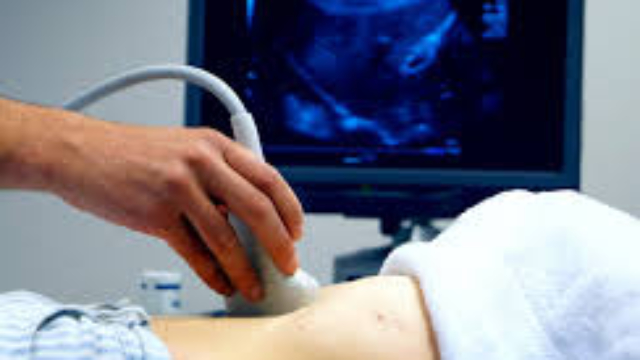
Ultrasound imaging starts with a device called a transducer. It sends high-frequency sound waves towards the body’s target area. This is the first step in creating diagnostic images.
The Ultrasound Procedure Explained
A sonographer uses the transducer during an ultrasound. They move it over the skin to get images from various angles. The sound waves reflect off internal structures and are picked up by the transducer.
The transducer sends this information to a computer. There, it’s processed to create the images we see.
What to Expect During an Examination
Patients usually lie on an examination table. A clear gel is applied to the skin to help sound waves pass through. The sonographer then moves the transducer over the area of interest.
The images appear on a monitor in real-time. This lets the sonographer see what they’re doing.
Preparation Guidelines
Preparation can vary for different ultrasound exams. Sometimes, patients need to fast or fill their bladder. Always follow the instructions from your healthcare provider or the ultrasound facility.
Interpreting Ultrasound Images
Radiologists, who are medical doctors, interpret the ultrasound images. They look for any abnormalities or conditions that need attention.
The Role of Sonographers and Radiologists
Sonographers are key in getting high-quality images. Radiologists use their knowledge to understand these images. Together, they make sure ultrasound exams give accurate and helpful information.
Medical Applications of Ultrasound
Ultrasound is very useful in many medical fields. It gives clear images in real-time without harmful radiation. This makes it perfect for diagnosing many health issues.
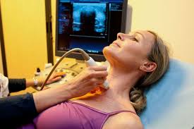
Obstetric and Gynecological Uses
Ultrasound is key in checking on babies during pregnancy. It helps spot any problems early. It also checks on women’s reproductive organs, finding issues like cysts or abnormalities.
Cardiovascular Imaging
Ultrasound is great for the heart. It checks how well the heart works and finds heart diseases. Echocardiography, a special ultrasound, shows the heart’s details, helping diagnose problems like valve issues.
Abdominal and Organ Scanning
Ultrasound looks at the liver, gallbladder, and kidneys. It finds gallstones, liver diseases, and other problems. It’s vital for diagnosing and treating stomach issues.
Musculoskeletal Applications
Ultrasound helps with muscle, tendon, and ligament injuries. It shows what’s happening in real-time. This helps doctors figure out and treat injuries like tendonitis or sprains.
Ultrasound’s wide use in medicine shows its value. It’s safe and gives clear images without radiation. This makes it a top choice for many medical tests.
Benefits of Ultrasound as a Radiation-Free Diagnostic Tool
Ultrasound technology has changed medical imaging for the better. It’s safe and doesn’t use radiation. This makes it very useful in many medical areas.
Safety Profile Compared to Other Imaging Methods
Ultrasound is very safe. It doesn’t use radiation like X-rays and CT scans do. Instead, it uses sound waves to see inside the body. This makes it perfect for people who are more sensitive.
Ultrasound vs. X-rays and CT Scans
Ultrasound is safer because it doesn’t use ionizing radiation. This is good for patients who need many scans or are pregnant. X-rays and CT scans use radiation, which can be a small risk.
Safety During Pregnancy
Ultrasound is safe for pregnant women. It’s used to check on the baby and find any problems. It doesn’t harm the mother or the baby.
Accessibility and Cost-Effectiveness
Ultrasound is not just safe but also easy to get and affordable. The machines are not very expensive. Plus, the test doesn’t need a lot of setup or time to recover. This makes it great for both patients and doctors.
In short, ultrasound is a great tool for doctors. It’s safe, easy to get, and doesn’t cost a lot. These reasons make it a top choice for many medical needs.
Limitations and Advancements in Ultrasound Technology
Ultrasound technology is always getting better, but it’s not perfect yet. Knowing what it can’t do helps us see how far it’s come and where it’s going.
Current Limitations of Ultrasound
Ultrasound has some big challenges. Two main ones are how clear the images can be and how much it depends on the person using it.
Image Quality Constraints
Ultrasound images can vary a lot. Things like the patient’s body type and the operator’s skill affect how clear the pictures are.
Operator Dependency
Ultrasound needs a skilled person to get good results. If the person doing it isn’t experienced, the images might not be as good.
Emerging Technologies and Future Directions
Ultrasound is getting better, thanks to new technologies. Things like artificial intelligence and portable devices are making it more useful.
Artificial Intelligence in Ultrasound
Artificial intelligence is making ultrasound images better. AI helps make pictures clearer and spot problems more easily.
Portable and Point-of-Care Ultrasound
New devices make ultrasound easier to use. They let doctors do scans right at the patient’s bedside, making care better.
As ultrasound tech keeps improving, it will likely overcome its current limits. This will make it even more useful for doctors and patients alike.
Ultrasound imaging is now a key tool in radiology. It’s safe and shows what’s inside without using harmful radiation.
This technology is used in many ways. It helps in checking on babies during pregnancy, looking at the heart, and scanning the abdomen. It’s also great for checking muscles and bones.
As ultrasound tech gets better, it will help more in healthcare. Doctors will have a powerful tool for finding and treating problems.
It’s important for doctors and patients to know about ultrasound imaging. It helps make better decisions and ensures the best care.
FAQ
What is ultrasound imaging?
Ultrasound imaging, also known as sonography, uses sound waves to see inside the body. It’s a safe way to look at organs and tissues without using harmful radiation. This method is non-invasive and helps doctors diagnose many conditions.
Is ultrasound imaging safe?
Yes, ultrasound imaging is safe. It doesn’t use harmful radiation, making it great for pregnant women. But, it should only be done by a trained healthcare professional.
What are the different types of ultrasound technologies?
There are many ultrasound technologies. 2D ultrasound shows two-dimensional images. 3D and 4D ultrasound create three-dimensional images and moving videos. Doppler ultrasound checks blood flow and finds vascular problems.
How do I prepare for an ultrasound examination?
Preparing for an ultrasound depends on the type of scan. For some, like abdominal scans, you might need to fast or avoid certain foods. Always follow the instructions from your healthcare provider or radiology department.
What is the role of sonographers and radiologists in ultrasound imaging?
Sonographers perform ultrasound exams and take images. Radiologists are doctors who read these images. Together, they help make accurate diagnoses and care plans.
Can ultrasound imaging be used during pregnancy?
Yes, ultrasound is often used during pregnancy. It helps track fetal growth, spot issues, and guide prenatal care. It’s safe for pregnant women.
What are the limitations of ultrasound imaging?
Ultrasound imaging is very useful but has some limits. Image quality can be affected by body type, gas in the bowel, and scar tissue. Also, the skill of the sonographer plays a big role in image quality.
Add a Comment