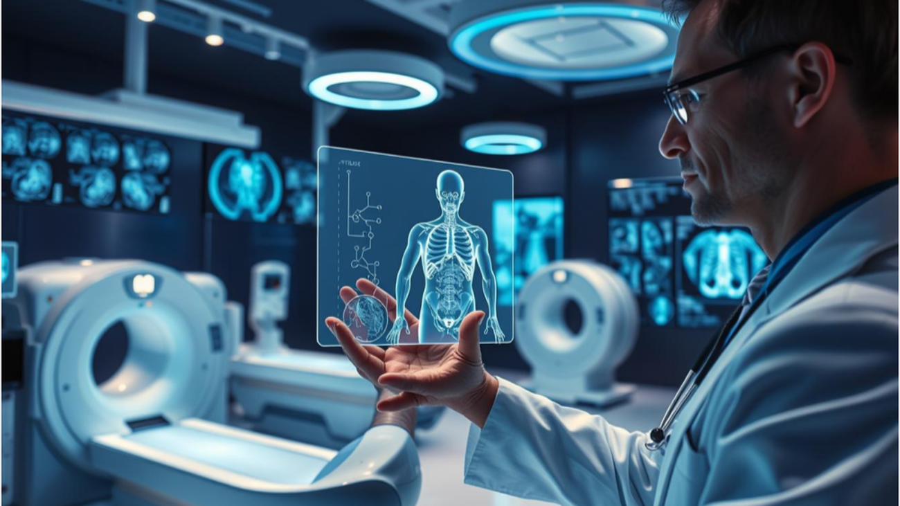
Medical diagnosis has seen a big change with AI-enhanced imaging. Now, doctors can use CT, MRI, and ultrasound together. This helps them make more accurate diagnoses.
By mixing these technologies with AI algorithms, doctors can spot complex patterns. This leads to smarter diagnoses. It also gives a better understanding of what’s going on with the patient.
So, doctors can create better treatment plans. This improves how well patients do. The mix of AI Imaging and medical knowledge is changing diagnostics.
The Current Landscape of Medical Imaging
Medical imaging has made big strides, but it’s not perfect yet. There are issues with how accurate it is and what it can do. Radiology is always getting better, aiming for more precise and reliable ways to diagnose.
Challenges in Traditional Diagnostic Imaging
Methods like CT scans, MRI, and ultrasound are key in medical diagnosis. But, they have their own problems.
Limitations of Single-Modality Approaches
Using just one imaging method can miss the mark. For example, ultrasound depends a lot on the person doing it. And CT scans might not show soft tissues well enough.
Diagnostic Accuracy Concerns
Getting a correct diagnosis is a big deal in medical imaging. Single-method approaches can lead to wrong or late diagnoses. This can really affect how well patients do.
The Need for Integrated Approaches
Using more than one imaging method can give a fuller picture of what’s going on inside the body.
Complementary Information from Multiple Sources
Putting together data from CT, MRI, and ultrasound can help doctors feel more sure and accurate in their diagnoses.
Evolution Toward Precision Medicine
This way of combining imaging is moving us toward precision medicine. It means treatments will be more tailored to each patient, based on detailed diagnostic info.
The move to using more imaging methods and combining them is changing radiology. It’s helping doctors make better choices and improving care for patients.
Understanding Individual Imaging Modalities
Advanced medical imaging techniques like CT, MRI, and ultrasound have changed how we diagnose diseases. Each method has its own way of working, benefits, and drawbacks.
Computed Tomography (CT): Principles and Limitations
CT scans use X-rays to make detailed images of the body’s inside. CT scans are very useful in emergency situations, like finding internal injuries or bleeding.
Radiation Exposure Considerations
One big problem with CT scans is the ionizing radiation they use. This can raise the risk of cancer. But, the benefits often outweigh the risks, mainly in urgent care.
Optimal Applications in Diagnosis
CT scans are best for finding tumors, fractures, and vascular diseases. They give quick and accurate results, which are key in emergency medicine.
Magnetic Resonance Imaging (MRI): Capabilities and Constraints
MRI uses strong magnetic fields and radio waves to make detailed images of the body’s inside. MRI is great at showing soft tissues, which is why it’s so valuable for diagnosing brain, spinal cord, and joint problems.
Soft Tissue Visualization Advantages
MRI’s ability to show soft tissues in detail is a big plus. It helps diagnose things like ligament tears, herniated discs, and some cancers.
Time and Cost Factors
One drawback of MRI is how long it takes. It’s also more expensive than CT scans or ultrasound.
Ultrasound Technology: Strengths and Weaknesses
Ultrasound uses sound waves to create images of the body’s inside. It’s a safe and non-invasive method that’s used a lot for diagnosis.
Real-Time Imaging Benefits
Ultrasound’s biggest advantage is its ability to show images in real-time. This is really helpful during things like biopsies or watching a fetus grow.
Operator Dependency Challenges
But, ultrasound depends on the person doing the scan. The quality of the images can vary a lot based on the operator’s skill.
The Rise of AI Imaging in Modern Medicine
Artificial Intelligence is now a big part of radiology. It helps improve how we analyze and understand medical images. This change is making diagnosis faster and more accurate.
AI-Enhanced Multi-Modality Imaging: Combining CT, MRI, and Ultrasound for Smarter Diagnoses
Machine Learning Algorithms in Image Analysis
Machine learning algorithms are key in medical image analysis. They come in two types: supervised and unsupervised learning.
Supervised Learning for Classification Tasks
Supervised learning uses labeled data to teach algorithms. It’s great for spotting specific health issues in images.
Unsupervised Learning for Pattern Recognition
Unsupervised learning finds patterns in data without labels. It uncovers new insights and oddities in medical images.
Deep Learning Applications in Radiology
Deep learning, like Convolutional Neural Networks (CNNs), is making waves in radiology.
Convolutional Neural Networks (CNNs)
CNNs excel at recognizing patterns in images. This skill is vital for image classification tasks.
Transfer Learning Approaches
Transfer learning refines pre-trained models for radiology. It boosts performance and cuts down on the need for lots of labeled data.
Computer Vision Techniques for Medical Images
Computer vision is being used more in medical imaging. It’s for tasks like segmenting images, extracting features, and finding anomalies.
Segmentation and Feature Extraction
Segmentation isolates specific parts of medical images. Feature extraction measures these parts’ characteristics.
Anomaly Detection Methods
Anomaly detection spots unusual things in images. It’s key for early diagnosis and planning treatments.
| Technique | Application | Benefit |
| Supervised Learning | Classification Tasks | High Accuracy |
| Unsupervised Learning | Pattern Recognition | Discovers New Insights |
| CNNs | Image Classification | Complex Pattern Recognition |
How AI Enhances Each Imaging Modality
Artificial intelligence is changing medical imaging. It’s making CT, MRI, and ultrasound better. AI algorithms are improving image quality and analysis.
AI-Powered CT Image Reconstruction and Analysis
AI is transforming CT imaging. It’s improving how images are made and analyzed. Noise reduction and image quality improvement are key areas where AI shines.
Noise Reduction and Image Quality Improvement
AI is making CT images clearer. It reduces noise, helping doctors spot small problems. This is vital for accurate diagnoses.
Automated Lesion Detection
AI can find lesions in CT scans automatically. This helps doctors spot issues fast and accurately.
MRI Enhancement Through Artificial Intelligence
AI is boosting MRI technology. It’s improving accelerated acquisition techniques and quantitative analysis capabilities.
Accelerated Acquisition Techniques
AI is making MRI scans faster. This makes them more comfortable for patients and more efficient for machines.
Quantitative Analysis Capabilities
AI also allows for detailed MRI analysis. It gives insights into tissue and pathology.
Ultrasound Interpretation with AI Assistance
In ultrasound, AI helps with real-time guidance systems and automated measurements and calculations.
Real-Time Guidance Systems
AI-assisted ultrasound systems offer real-time feedback. This boosts procedure accuracy.
Automated Measurements and Calculations
AI also automates ultrasound tasks. This saves healthcare professionals time and reduces errors.
Multi-Modality Integration: Technical Foundations
Multi-modality imaging combines different techniques like CT, MRI, and ultrasound. This gives a full view of what’s going on inside the body. It’s all thanks to advanced tech that lets data from various sources come together.
Data Fusion Techniques for Medical Images
Data fusion is key for mixing info from different imaging types. Early fusion and late fusion are the main methods used in medical imaging.
Early Fusion vs. Late Fusion Approaches
Early fusion blends data at the image level. Late fusion, on the other hand, combines the results of each modality’s analysis. The choice depends on the clinical need.
Multiparametric Analysis Methods
Multiparametric analysis looks at many features from different imaging types. This method boosts accuracy by giving a detailed look at a patient’s health.
Co-registration and Alignment Challenges
Aligning images from different sources is vital for accurate fusion. Rigid and non-rigid registration algorithms help match images.
Rigid and Non-rigid Registration Algorithms
Rigid registration works for images that have only moved slightly. Non-rigid registration is for images that have changed shape.
Temporal Alignment Considerations
Temporal alignment is key for dynamic imaging. It makes sure images from different times match up.
Creating Comprehensive 3D Visualizations
Advanced visualization creates detailed 3D views of the combined data. Volume rendering techniques and interactive visualization tools help doctors understand complex images better.
Volume Rendering Techniques
Volume rendering makes 3D images from 3D data. It shows what’s inside the body.
Interactive Visualization Tools
Interactive tools let doctors play with 3D images. They can look at data from all sides, helping with diagnosis.
Clinical Applications of AI-Enhanced Multi-Modality Imaging
AI-enhanced multi-modality imaging has many uses in medicine. It helps in several key areas of medical practice. This technology is used in many medical fields to improve diagnosis and treatment.
Oncology: Tumor Detection and Characterization
In oncology, AI plays a big role in finding and understanding tumors. By mixing data from CT, MRI, and ultrasound, AI gives more accurate diagnoses.
Improved Staging and Treatment Planning
AI imaging helps in precise tumor staging. This is key for choosing the best treatment. For example, CT and PET data together show how far tumors have spread.
Monitoring Treatment Response
AI can track changes in tumors over time. This lets doctors see if treatments are working early on.
Neurology: Brain Mapping and Disorder Diagnosis
In neurology, AI helps map the brain and diagnose disorders. MRI is very useful here.
Stroke Assessment and Management
AI imaging quickly checks how severe a stroke is. It also finds the best treatment.
Neurodegenerative Disease Evaluation
Cardiovascular Applications: Structural and Functional Assessment
In heart medicine, AI imaging checks the heart’s structure and function.
Coronary Artery Disease Evaluation
CT angiography with AI checks coronary artery disease.
Cardiac Function Analysis
AI analyzes ultrasound and MRI data to check heart function.
Musculoskeletal Imaging: Improving Precision and Accuracy
In musculoskeletal imaging, AI makes diagnoses more precise and accurate.
Joint and Soft Tissue Assessment
MRI and ultrasound together check joints and soft tissues.
Trauma and Degenerative Disease Evaluation
AI imaging helps evaluate trauma and degenerative diseases.
| Medical Specialty | Primary Imaging Modalities | AI-Enhanced Applications |
| Oncology | CT, MRI, Ultrasound | Tumor detection, staging, treatment response monitoring |
| Neurology | MRI | Brain mapping, stroke assessment, neurodegenerative disease evaluation |
| Cardiovascular | CT, Ultrasound, MRI | Coronary artery disease evaluation, cardiac function analysis |
| Musculoskeletal | MRI, Ultrasound | Joint and soft tissue assessment, trauma and degenerative disease evaluation |
Benefits and Outcomes of Integrated Imaging Approaches
AI-powered imaging is changing how we diagnose and treat patients. It combines CT, MRI, and ultrasound to give doctors a clearer picture. This leads to more accurate and detailed diagnoses.
Improved Diagnostic Accuracy and Confidence
AI makes diagnoses better by analyzing complex data. AI algorithms can spot patterns that humans might miss.
Reduction in False Positives and Negatives
AI helps avoid wrong diagnoses. This is key in radiology, where getting it right is critical.
Enhanced Lesion Characterization
AI can better understand lesions. It gives detailed info on their nature and size. This helps doctors plan treatments.
Reduced Need for Invasive Procedures
AI reduces the need for risky tests. Non-invasive tissue analysis makes exams safer and more comfortable for patients.
Non-invasive Tissue Characterization
AI supports detailed tissue analysis without biopsies. This is a big step forward in diagnostics.
Virtual Biopsy Concepts
AI is making virtual biopsies possible. This could change how we diagnose diseases, making it safer and more precise.
Time and Cost Efficiency in the Diagnostic Process
AI speeds up and saves money in diagnosis. It makes the process more efficient and cost-effective.
Streamlined Workflow Integration
AI fits into current workflows, boosting efficiency. This helps doctors work better and faster.
Resource Optimization Strategies
AI helps use resources wisely. This cuts costs and improves patient care.
Patient-Centered Advantages
AI imaging benefits patients in many ways. It offers personalized care and better experiences.
Personalized Diagnostic Approaches
AI tailors care to each patient. This improves the quality of care for everyone.
Improved Patient Experience
AI reduces the need for risky tests. This makes the diagnostic process smoother and safer for patients.
Implementation Challenges and Considerations
Using AI in multi-modality imaging, like CT and ultrasound, is not easy. It needs a big effort to get past technical, regulatory, and educational barriers.
Technical Infrastructure Requirements
To support AI in imaging, you need a lot of tech. This includes fast computers and lots of storage for big data.
Computing Resources and Storage Needs
Advanced tech is key for handling complex data. High-capacity storage solutions are also needed to store all the data.
Network and Integration Considerations
Connecting different imaging types and AI needs a strong network. It’s important for systems to work well together.
Regulatory and Approval Pathways
Getting AI imaging approved is a big challenge. You must follow rules to use it in clinics.
FDA and Health Canada Clearance Processes
Knowing the rules from the FDA and Health Canada is important. These rules make sure AI imaging is safe and works well.
Validation Requirements for Clinical Use
AI and imaging systems must be tested well. This shows they are useful and accurate in clinics.
Training Requirements for Healthcare Professionals
Healthcare workers need good training for AI imaging. They must learn how to use and understand AI insights.
Radiologist Education and Adaptation
Radiologists and others need to learn about AI tools. They must know what AI can do and what it can’t.
Multidisciplinary Team Collaboration
Working together is key for AI imaging success. Teams of experts are needed to make the most of AI.
Ethical and Privacy Considerations
AI in imaging raises big questions about ethics and privacy. Keeping patient data safe is very important.
Data Security and Patient Confidentiality
Protecting patient info is a must. Following privacy rules is essential.
Algorithmic Transparency and Bias Mitigation
AI tools must be clear and fair. Regular checks and updates help keep trust in AI.
AI is changing how we do medical imaging, like MRI and other radiology methods. By using CT, MRI, and ultrasound together, doctors can understand patients better. This leads to more accurate diagnoses and treatments.
AI algorithms are getting better, making radiology more precise. This means doctors can analyze complex images with great accuracy. The future of AI in imaging looks very promising for better patient care and easier clinical work.
To make the most of AI in imaging, we need to keep investing in technology, rules, and training doctors. With AI, the medical field is set for a new era of excellence in diagnosis and care.
FAQ
What is AI-enhanced multi-modality imaging?
AI-enhanced multi-modality imaging uses different medical imaging tech like CT, MRI, and ultrasound. It adds artificial intelligence to boost how well doctors can diagnose and feel sure about their findings.
How does AI enhance medical imaging modalities like CT, MRI, and ultrasound?
AI makes these imaging methods better by improving how images are made, analyzed, and understood. For instance, AI can clean up CT images, speed up MRI scans, and guide ultrasound procedures in real-time.
What are the benefits of using AI-enhanced multi-modality imaging in clinical practice?
Using AI in imaging helps doctors make more accurate diagnoses. It also means fewer invasive tests, better patient experiences, and a smoother workflow. All these lead to better health outcomes for patients.
What are the challenges in implementing AI-enhanced multi-modality imaging?
There are a few hurdles to overcome. These include setting up the right tech, getting approvals, training staff, and thinking about ethics like keeping patient data safe and being open about how AI works.
How does AI-enhanced multi-modality imaging impact patient care in oncology?
In cancer care, AI helps spot tumors better and understand their size and type. It also helps plan treatments and check how well they’re working. This leads to more tailored and effective cancer care.
What role does radiology play in AI-enhanced multi-modality imaging?
Radiology is key in using AI for imaging. Radiologists look at images and work with AI to make diagnoses more accurate and confident.
Are there any specific regulations or guidelines for the use of AI in medical imaging in Canada?
Yes, Health Canada and other groups have rules and checks for AI in imaging. These ensure it’s safe and works well.
How does AI-enhanced multi-modality imaging contribute to precision medicine?
AI imaging helps precision medicine by giving detailed, personal info for diagnosis. This allows for targeted treatments and better health results for patients.
Add a Comment